Application
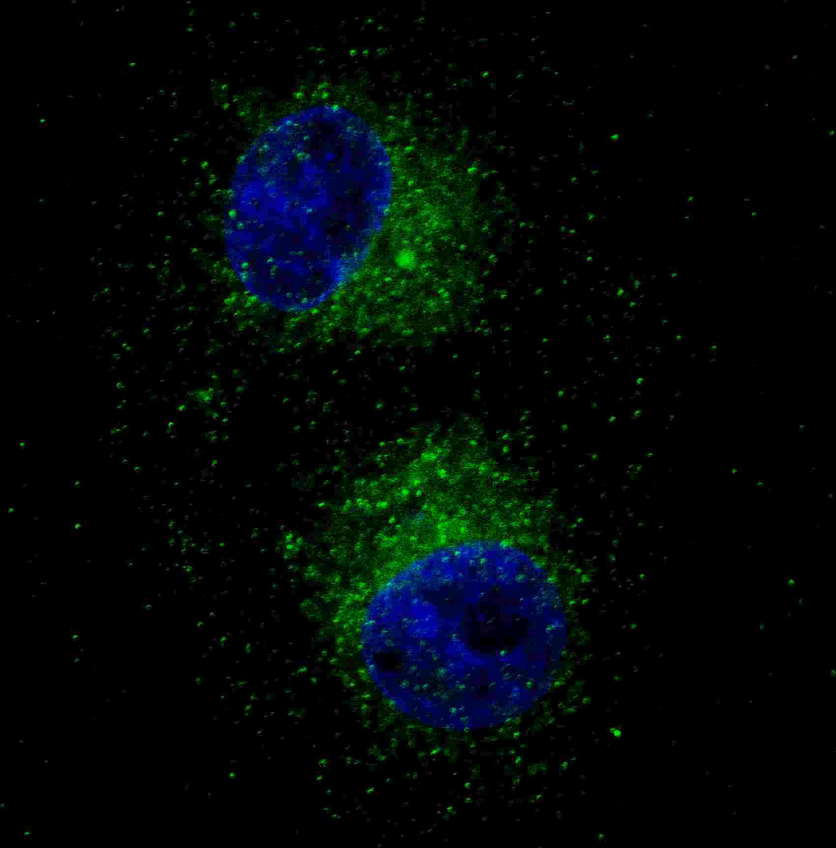
Fluorescent confocal image of HepG2 cells stained with MET/HGFR antibody. HepG2 cells were fixed with 4% PFA (20 min), permeabilized with Triton X-100 (0.2%, 30 min). Cells were then incubated with AM1002a MET/HGFR primary antibody (1:100, 2 h at room temperature). For secondary antibody, Alexa Fluor® 488 conjugated donkey anti-mouse antibody (green) was used (1:1000, 1h). Nuclei were counterstained with Hoechst 33342 (blue) (10 μg/ml, 5 min). ). Note the highly specific localization of the MET immunosignal to the cytoplasm, supported by Human Protein Atlas Data (http://www.proteinatlas.org/ENSG00000105976).Application
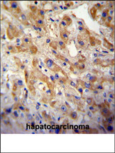
MET/HGFR Antibody (Cat. #AM1002a) immunohistochemistry analysis in formalin fixed and paraffin embedded human hepatocarcinoma followed by peroxidase conjugation of the secondary antibody and DAB staining.This data demonstrates the use of the MET/HGFR Antibody for immunohistochemistry. Clinical relevance has not been evaluated.Application
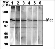
Detection of endogenous Met in HepG2 cell line. 10 ug/lane of HepG2 cell lysate was used to examine the expression of human Met. Lanes 1-5 represent Abgent s different anti-Met monoclonal antibodies that are Cat# AM1001-1005. Lane 6 represents auto-phosohorylated-Met in HepG2 cell line detected by anti-phospho-Met Mab (Cat# AM1000).| Product Name | MET/HGFR Antibody |
|---|---|
| Antibody Type | Primary Antibodies |
| Antigen Alias | MET; Hepatocyte growth factor receptor; HGF/SF receptor; Proto-oncogene c-Met; Scatter factor receptor; Tyrosine-protein kinase Met |
| Clonality | Monoclonal |
|---|---|
| Isotype | IgG1 |
| Clone Number | 6AT203 |
| Host Species | Mouse |
| Tested Applications | IFIHCWB |
| IF:1:100 IHC |
|
| Species Reactivity | Human |
| Concentration | 1mg/ml |
| Alternative Names | MET Hepatocyte growth factor receptor HGF/SF receptor Proto-oncogene c-Met Scatter factor receptor Tyrosine-protein kinase Met |
|---|---|
| Molecular Weight(MW) | 155541 Da |
| Function | Receptor tyrosine kinase that transduces signals from the extracellular matrix into the cytoplasm by binding to hepatocyte growth factor/HGF ligand. Regulates many physiological processes including proliferation, scattering, morphogenesis and survival. Ligand binding at the cell surface induces autophosphorylation of MET on its intracellular domain that provides docking sites for downstream signaling molecules Following activation by ligand, interacts with the PI3-kinase subunit PIK3R1, PLCG1, SRC, GRB2, STAT3 or the adapter GAB1 Recruitment of these downstream effectors by MET leads to the activation of several signaling cascades including the RAS-ERK, PI3 kinase-AKT, or PLCgamma-PKC. The RAS-ERK activation is associated with the morphogenetic effects while PI3K/AKT coordinates prosurvival effects. During embryonic development, MET signaling plays a role in gastrulation, development and migration of muscles and neuronal precursors, angiogenesis and kidney formation. In adults, participates in wound healing as well as organ regeneration and tissue remodeling. Promotes also differentiation and proliferation of hematopoietic cells |
| Tissue Specificity | Expressed in normal hepatocytes as well as in epithelial cells lining the stomach, the small and the large intestine. Found also in basal keratinocytes of esophagus and skin. High levels are found in liver, gastrointestinal tract, thyroid and kidney. Also present in the brain |
| Cellular Localization | Membrane; Single-pass type I membrane protein |
| Entrez Gene | 4233 |
|---|
Application
Fluorescent confocal image of HepG2 cells stained with MET/HGFR antibody. HepG2 cells were fixed with 4% PFA (20 min), permeabilized with Triton X-100 (0.2%, 30 min). Cells were then incubated with AM1002a MET/HGFR primary antibody (1:100, 2 h at room temperature). For secondary antibody, Alexa Fluor® 488 conjugated donkey anti-mouse antibody (green) was used (1:1000, 1h). Nuclei were counterstained with Hoechst 33342 (blue) (10 μg/ml, 5 min). ). Note the highly specific localization of the MET immunosignal to the cytoplasm, supported by Human Protein Atlas Data (http://www.proteinatlas.org/ENSG00000105976).
Application
MET/HGFR Antibody (Cat. #AM1002a) immunohistochemistry analysis in formalin fixed and paraffin embedded human hepatocarcinoma followed by peroxidase conjugation of the secondary antibody and DAB staining.This data demonstrates the use of the MET/HGFR Antibody for immunohistochemistry. Clinical relevance has not been evaluated.
Application
Detection of endogenous Met in HepG2 cell line. 10 ug/lane of HepG2 cell lysate was used to examine the expression of human Met. Lanes 1-5 represent Abgent s different anti-Met monoclonal antibodies that are Cat# AM1001-1005. Lane 6 represents auto-phosohorylated-Met in HepG2 cell line detected by anti-phospho-Met Mab (Cat# AM1000).| Application Notes | IF:1:100 IHC |
|---|
| Form | Liquid |
|---|---|
| Storage Instructions | For short-term storage, store at 4° C. For long-term storage, aliquot and store at -20ºC or below. Avoid multiple freeze-thaw cycles. |
| Storage Buffer | Purified monoclonal antibody supplied in PBS with 0.09% (W/V) sodium azide. This antibody is purified through a protein G column, eluted with high and low pH buffers and neutralized immediately, followed by dialysis against PBS. |


