Application
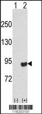
Western blot analysis of MLH1 (arrow) using rabbit polyclonal MLH1 Antibody (Center) . 293 cell lysates (2 ug/lane) either nontransfected (Lane 1) or transiently transfected with the MLH1 gene (Lane 2) .Application
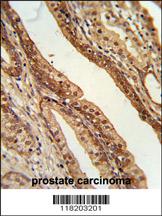
Formalin-fixed and paraffin-embedded human prostate carcinoma reacted with MLH1 Antibody (Center), which was peroxidase-conjugated to the secondary antibody, followed by DAB staining. This data demonstrates the use of this antibody for immunohistochemistry; clinical relevance has not been evaluated.Application
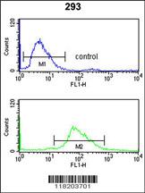
MLH1 Antibody (Center) flow cytometry analysis of 293 cells (bottom histogram) compared to a negative control cell (top histogram).FITC-conjugated goat-anti-rabbit secondary antibodies were used for the analysis.Application
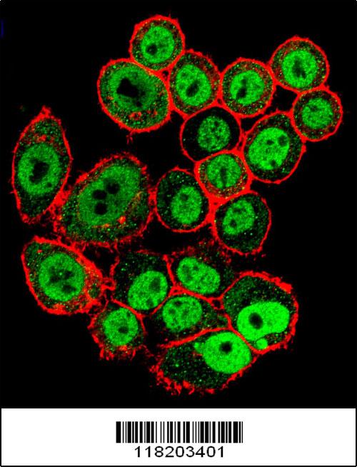
Confocal immunofluorescent analysis of MLH1 Antibody (Center)(Cat#AP7464c) with Hela cell followed by Alexa Fluor 488-conjugated goat anti-rabbit lgG (green). Actin filaments have been labeled with Alexa Fluor 555 phalloidin (red).| Product Name | MLH1 Antibody (Center) |
|---|---|
| Antibody Type | Primary Antibodies |
| Antigen Alias | MLH1; COCA2; DNA mismatch repair protein Mlh1; MutL protein homolog 1 |
| Clonality | Polyclonal |
|---|---|
| Isotype | Ig |
| Host Species | Rabbit |
| Tested Applications | WBIHCFCIF |
| WB:1:100~500 IHC |
|
| Species Reactivity | Human |
| Concentration | 1mg/ml |
| Gene Synonyms | COCA2 |
|---|---|
| Alternative Names | MLH1 COCA2 DNA mismatch repair protein Mlh1 MutL protein homolog 1 |
| Molecular Weight(MW) | 84601 Da |
| Function | Heterodimerizes with PMS2 to form MutL alpha, a component of the post-replicative DNA mismatch repair system (MMR). DNA repair is initiated by MutS alpha (MSH2-MSH6) or MutS beta (MSH2-MSH6) binding to a dsDNA mismatch, then MutL alpha is recruited to the heteroduplex. Assembly of the MutL-MutS- heteroduplex ternary complex in presence of RFC and PCNA is sufficient to activate endonuclease activity of PMS2. It introduces single-strand breaks near the mismatch and thus generates new entry points for the exonuclease EXO1 to degrade the strand containing the mismatch. DNA methylation would prevent cleavage and therefore assure that only the newly mutated DNA strand is going to be corrected. MutL alpha (MLH1-PMS2) interacts physically with the clamp loader subunits of DNA polymerase III, suggesting that it may play a role to recruit the DNA polymerase III to the site of the MMR. Also implicated in DNA damage signaling, a process which induces cell cycle arrest and can lead to apoptosis in case of major DNA damages. Heterodimerizes with MLH3 to form MutL gamma which plays a role in meiosis |
| Tissue Specificity | Colon, lymphocytes, breast, lung, spleen, testis, prostate, thyroid, gall bladder and heart |
| Cellular Localization | Nucleus. |
| Entrez Gene | 4292 |
|---|
Application
Western blot analysis of MLH1 (arrow) using rabbit polyclonal MLH1 Antibody (Center) . 293 cell lysates (2 ug/lane) either nontransfected (Lane 1) or transiently transfected with the MLH1 gene (Lane 2) .
Application
Formalin-fixed and paraffin-embedded human prostate carcinoma reacted with MLH1 Antibody (Center), which was peroxidase-conjugated to the secondary antibody, followed by DAB staining. This data demonstrates the use of this antibody for immunohistochemistry; clinical relevance has not been evaluated.
Application
MLH1 Antibody (Center) flow cytometry analysis of 293 cells (bottom histogram) compared to a negative control cell (top histogram).FITC-conjugated goat-anti-rabbit secondary antibodies were used for the analysis.
Application
Confocal immunofluorescent analysis of MLH1 Antibody (Center)(Cat#AP7464c) with Hela cell followed by Alexa Fluor 488-conjugated goat anti-rabbit lgG (green). Actin filaments have been labeled with Alexa Fluor 555 phalloidin (red).| Application Notes | WB:1:100~500 IHC |
|---|
| Form | Liquid |
|---|---|
| Storage Instructions | For short-term storage, store at 4° C. For long-term storage, aliquot and store at -20ºC or below. Avoid multiple freeze-thaw cycles. |
| Storage Buffer | Purified polyclonal antibody supplied in PBS with 0.09% (W/V) sodium azide. This antibody is purified through a protein A column, followed by peptide affinity purification. |


