Application
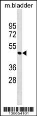
Mouse Parva Antibody (N-term) western blot analysis in mouse bladder tissue lysates (35ug/lane).This demonstrates the Parva antibody detected the Parva protein (arrow).| Product Name | Parva Antibody (N-term) |
|---|---|
| Antibody Type | Primary Antibodies |
| Clonality | Polyclonal |
|---|---|
| Isotype | Ig |
| Host Species | Rabbit |
| Tested Applications | WB |
| WB:1:100~500 |
|
| Species Reactivity | Human |
| Concentration | 1mg/ml |
| Gene Synonyms | Actp |
|---|---|
| Alternative Names | Parva Actp Alpha-parvin Actopaxin |
| Molecular Weight(MW) | 42330 Da |
| Function | Plays a role in the reorganization of the actin cytoskeleton, formation of lamellipodia and ciliogenesis. Plays a role in the establishement of cell polarity, cell adhesion, cell spreading, and directed cell migration. Plays a role in sarcomere organization and in smooth muscle cell contraction. Required for normal development of the embryonic cardiovascular system, and for normal septation of the heart outflow tract. Plays a role in sprouting angiogenesis and is required for normal adhesion of vascular smooth muscle cells to endothelial cells during blood vessel development |
| Tissue Specificity | This Mouse Parva antibody is generated from rabbits immunized with a KLH conjugated synthetic peptide between 65-93 amino acids from the N-terminal region of mouse Parva. |
| Cellular Localization | Cell junction, focal adhesion. Cell membrane; Peripheral membrane protein; Cytoplasmic side Cytoplasm, cytoskeleton. Cytoplasm, myofibril, sarcomere, Z line Note=Constituent of focal adhesions |
| Entrez Gene | 57342 |
|---|---|
| Nucleotide Accession | NP_065631.3 |
Application
Mouse Parva Antibody (N-term) western blot analysis in mouse bladder tissue lysates (35ug/lane).This demonstrates the Parva antibody detected the Parva protein (arrow).| Application Notes | WB:1:100~500 |
|---|
| Form | Liquid |
|---|---|
| Storage Instructions | For short-term storage, store at 4° C. For long-term storage, aliquot and store at -20ºC or below. Avoid multiple freeze-thaw cycles. |
| Storage Buffer | Purified polyclonal antibody supplied in PBS with 0.09% (W/V) sodium azide. This antibody is purified through a protein A column, followed by peptide affinity purification. |


