Application
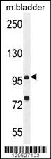
NOD1 Antibody (C-term) western blot analysis in mouse bladder tissue lysates (35ug/lane).This demonstrates the NOD1 antibody detected the NOD1 protein (arrow).Application
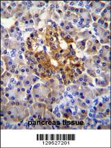
NOD1 Antibody (C-term) immunohistochemistry analysis in formalin fixed and paraffin embedded human pancreas tissue followed by peroxidase conjugation of the secondary antibody and DAB staining.This data demonstrates the use of NOD1 Antibody (C-term) for immunohistochemistry. Clinical relevance has not been evaluated.Application
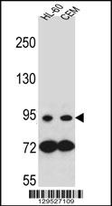
NOD1 Antibody (C-term) western blot analysis in HL-60,CEM cell line lysates (35ug/lane).This demonstrates the NOD1 antibody detected the NOD1 protein (arrow).Application
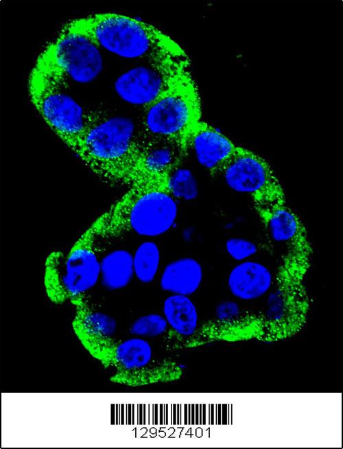
Confocal immunofluorescent analysis of NOD1 Antibody (C-term)(Cat#AP11805b) with HepG2 cell followed by Alexa Fluor 488-conjugated goat anti-rabbit lgG (green). DAPI was used to stain the cell nuclear (blue).Application
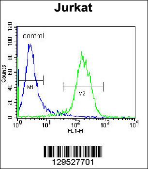
NOD1 Antibody (C-term) flow cytometric analysis of Jurkat cells (right histogram) compared to a negative control cell (left histogram).FITC-conjugated donkey-anti-rabbit secondary antibodies were used for the analysis.| Product Name | NOD1 Antibody (C-term) |
|---|---|
| Antibody Type | Primary Antibodies |
| Clonality | Polyclonal |
|---|---|
| Isotype | Ig |
| Host Species | Rabbit |
| Tested Applications | WBIHCIFFC |
| WB:1:100~500 IHC |
|
| Species Reactivity | Human |
| Concentration | 1mg/ml |
| Gene Synonyms | CARD4 |
|---|---|
| Alternative Names | NOD1 CARD4 Nucleotide-binding oligomerization domain-containing protein 1 Caspase recruitment domain-containing protein 4 |
| Molecular Weight(MW) | 107691 Da |
| Function | Enhances caspase-9-mediated apoptosis. Induces NF-kappa- B activity via RIPK2 and IKK-gamma. Confers responsiveness to intracellular bacterial lipopolysaccharides (LPS). Forms an intracellular sensing system along with ARHGEF2 for the detection of microbial effectors during cell invasion by pathogens. Required for RHOA and RIPK2 dependent NF-kappa-B signaling pathway activation upon S.flexneri cell invasion. Involved not only in sensing peptidoglycan (PGN)-derived muropeptides but also in the activation of NF-kappa-B by Shigella effector proteins IpgB2 and OspB. Recruits NLRP10 to the cell membrane following bacterial infection |
| Tissue Specificity | Highly expressed in adult heart, skeletal muscle, pancreas, spleen and ovary. Also detected in placenta, lung, liver, kidney, thymus, testis, small intestine and colon |
| Cellular Localization | Cytoplasm. Cell membrane. Apical cell membrane. Basolateral cell membrane. Note=Detected in the cytoplasm and at the cell membrane. Following bacterial infection, localizes to bacterial entry sites in the cell membrane. Recruited to the basolateral and apical membranes in polarized epithelial cells |
| Entrez Gene | 10392 |
|---|---|
| Nucleotide Accession | NP_006083.1 |
Application
NOD1 Antibody (C-term) western blot analysis in mouse bladder tissue lysates (35ug/lane).This demonstrates the NOD1 antibody detected the NOD1 protein (arrow).
Application
NOD1 Antibody (C-term) immunohistochemistry analysis in formalin fixed and paraffin embedded human pancreas tissue followed by peroxidase conjugation of the secondary antibody and DAB staining.This data demonstrates the use of NOD1 Antibody (C-term) for immunohistochemistry. Clinical relevance has not been evaluated.
Application
NOD1 Antibody (C-term) western blot analysis in HL-60,CEM cell line lysates (35ug/lane).This demonstrates the NOD1 antibody detected the NOD1 protein (arrow).
Application
Confocal immunofluorescent analysis of NOD1 Antibody (C-term)(Cat#AP11805b) with HepG2 cell followed by Alexa Fluor 488-conjugated goat anti-rabbit lgG (green). DAPI was used to stain the cell nuclear (blue).
Application
NOD1 Antibody (C-term) flow cytometric analysis of Jurkat cells (right histogram) compared to a negative control cell (left histogram).FITC-conjugated donkey-anti-rabbit secondary antibodies were used for the analysis.| Application Notes | WB:1:100~500 IHC |
|---|
| Form | Liquid |
|---|---|
| Storage Instructions | For short-term storage, store at 4° C. For long-term storage, aliquot and store at -20ºC or below. Avoid multiple freeze-thaw cycles. |
| Storage Buffer | Purified polyclonal antibody supplied in PBS with 0.09% (W/V) sodium azide. This antibody is purified through a protein A column, followed by peptide affinity purification. |


