WB
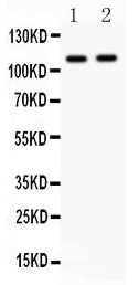
Western blot analysis of CARD4/NOD1 using anti-CARD4/NOD1 antibody. The sample well of each lane was loaded with 30 ug of sample under reducing conditions. Lane 1: A549 whole cell lysates, Lane 2: Rat Cardiac Muscle tissue lysates. After electrophoresis, proteins were transferred to a membrane. Then the membrane was incubated with rabbit anti-CARD4/NOD1 antigen affinity purified polyclonal antibody at a dilution of 1:1000 and probed with a goat anti-rabbit IgG-HRP secondary antibody . The signal is developed using ECL Plus Western Blotting Substrate.IHC
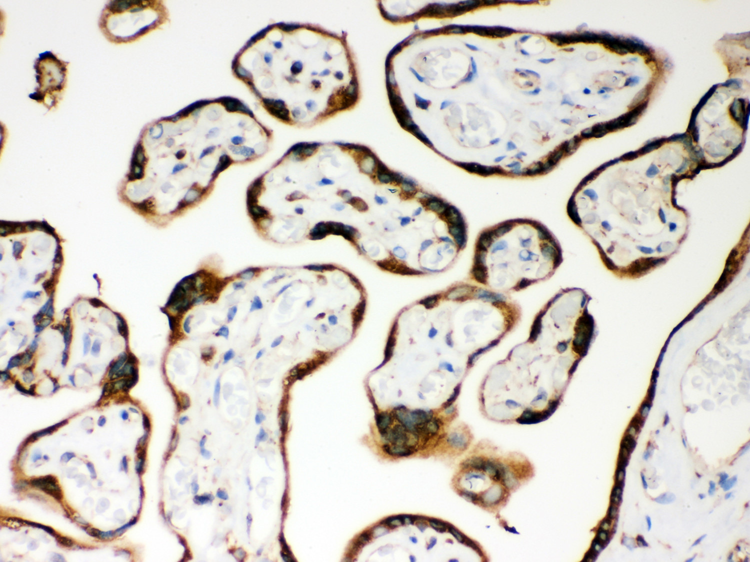
IHC analysis of CARD4/NOD1 using anti-CARD4/NOD1 antibody. CARD4/NOD1 was detected in a paraffin-embedded section of human placenta tissue. Biotinylated goat anti-rabbit IgG was used as secondary antibody. The tissue section was incubated with rabbit anti-CARD4/NOD1 Antibody at a dilution of 1:200 and developed using Strepavidin-Biotin-Complex (SABC) with DAB as the chromogen.ICC/IF
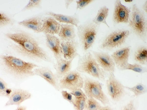
ICC analysis of CARD4/NOD1 using anti- CARD4/NOD1 antibody. CARD4/NOD1 was detected in an immunocytochemical section of A549 cells. The section was incubated with rabbit anti-CARD4/NOD1 Antibody at a dilution of 1:100. Biotinylated goat anti-rabbit IgG was used as secondary antibody. The section was developed using Strepavidin-Biotin-Complex (SABC) with DAB as the chromogen.| Product Name | Rabbit polyclonal antibody to CARD4/NOD1 |
|---|---|
| Antibody Type | Primary Antibodies |
| Immunogen | E.coli-derived human CARD4 recombinant protein (Position: M1-M160). Human CARD4 shares 82% amino acid (aa) sequence identity with mouse CARD4. |
| Clonality | Polyclonal |
|---|---|
| Isotype | IgG |
| Host Species | Rabbit |
| Tested Applications | ICC/IFIHCWB |
| WB:1:500-1:2000 IHC:1:50-1:400 ICC/IF:1:50-1:400 |
|
| Species Reactivity | HumanRat |
| Concentration | 0.5mg/ml |
| Purification | Affinity purified |
| Gene Symbol | NOD1 |
|---|---|
| Gene Synonyms | CARD4 NLRC1 hNod1 CLR7.1 |
| Gene Full Name | nucleotide binding oligomerization domain containing 1 |
| Gene Summary | This gene encodes a member of the nucleotide-binding oligomerization domain (NOD)-like receptor (NLR) family of proteins. The encoded protein plays a role in innate immunity by acting as a pattern-recognition receptor (PRR) that binds bacterial peptidoglycans and initiates inflammation. This protein has also been implicated in the immune response to viral and parasitic infection. Major structural features of this protein include an N-terminal caspase recruitment domain (CARD), a centrally located nucleotide-binding domain (NBD), and 10 tandem leucine-rich repeats (LRRs) in its C terminus. The CARD is involved in apoptotic signaling, LRRs participate in protein-protein interactions, and mutations in the NBD may affect the process of oligomerization and subsequent function of the LRR domain. Mutations in this gene are associated with asthma, inflammatory bowel disease, Behcet disease and sarcoidosis in human patients. [provided by RefSeq, Aug 2017] |
| Alternative Names | nucleotide binding oligomerization domain containing 1 CARD4 CLR7.1 NLRC1 NOD1 |
| Molecular Weight(MW) | 108 kDa(Observed band size:107 kDa) |
| Cellular Localization | Cell membrane,Cytoplasm,Membrane. |
WB
Western blot analysis of CARD4/NOD1 using anti-CARD4/NOD1 antibody. The sample well of each lane was loaded with 30 ug of sample under reducing conditions. Lane 1: A549 whole cell lysates, Lane 2: Rat Cardiac Muscle tissue lysates. After electrophoresis, proteins were transferred to a membrane. Then the membrane was incubated with rabbit anti-CARD4/NOD1 antigen affinity purified polyclonal antibody at a dilution of 1:1000 and probed with a goat anti-rabbit IgG-HRP secondary antibody . The signal is developed using ECL Plus Western Blotting Substrate.
IHC
IHC analysis of CARD4/NOD1 using anti-CARD4/NOD1 antibody. CARD4/NOD1 was detected in a paraffin-embedded section of human placenta tissue. Biotinylated goat anti-rabbit IgG was used as secondary antibody. The tissue section was incubated with rabbit anti-CARD4/NOD1 Antibody at a dilution of 1:200 and developed using Strepavidin-Biotin-Complex (SABC) with DAB as the chromogen.
ICC/IF
ICC analysis of CARD4/NOD1 using anti- CARD4/NOD1 antibody. CARD4/NOD1 was detected in an immunocytochemical section of A549 cells. The section was incubated with rabbit anti-CARD4/NOD1 Antibody at a dilution of 1:100. Biotinylated goat anti-rabbit IgG was used as secondary antibody. The section was developed using Strepavidin-Biotin-Complex (SABC) with DAB as the chromogen.| Application Notes | WB:1:500-1:2000 IHC:1:50-1:400 ICC/IF:1:50-1:400 |
|---|
| Form | Liquid |
|---|---|
| Storage Instructions | 12 months from date of receipt,-20℃ as supplied. |
| Storage Buffer | 500 ug/ml antibody with PBS, 0.02% NaN3, 1 mg/ml BSA and 50% glycerol. |


