Application
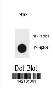
Dot blot analysis of p27Kip1 Antibody (Phospho S83) Phospho-specific Pab on nitrocellulose membrane. 50ng of Phospho-peptide or Non Phospho-peptide per dot were adsorbed. Antibody working concentrations are 0.6ug per ml.| Product Name | p27Kip1 Antibody (Phospho S83) |
|---|---|
| Antibody Type | Primary Antibodies |
| Antigen Alias | CDKN1B; KIP1; Cyclin-dependent kinase inhibitor 1B; Cyclin-dependent kinase inhibitor p27; p27Kip1 |
| Modification | p-S83 |
| Clonality | Polyclonal |
|---|---|
| Isotype | Ig |
| Host Species | Rabbit |
| Tested Applications | DB |
| DB:1:500 |
|
| Species Reactivity | Human |
| Concentration | 1mg/ml |
| Gene Synonyms | KIP1 |
|---|---|
| Alternative Names | CDKN1B KIP1 Cyclin-dependent kinase inhibitor 1B Cyclin-dependent kinase inhibitor p27 p27Kip1 |
| Molecular Weight(MW) | 22073 Da |
| Function | Important regulator of cell cycle progression. Involved in G1 arrest. Potent inhibitor of cyclin E- and cyclin A-CDK2 complexes. Forms a complex with cyclin type D-CDK4 complexes and is involved in the assembly, stability, and modulation of CCND1- CDK4 complex activation. Acts either as an inhibitor or an activator of cyclin type D-CDK4 complexes depending on its phosphorylation state and/or stoichometry |
| Tissue Specificity | Expressed in all tissues tested. Highest levels in skeletal muscle, lowest in liver and kidney |
| Cellular Localization | Nucleus. Cytoplasm. Endosome (By similarity). Note=Nuclear and cytoplasmic in quiescent cells. AKT- or RSK-mediated phosphorylation on Thr-198, binds 14-3-3, translocates to the cytoplasm and promotes cell cycle progression Mitogen-activated UHMK1 phosphorylation on Ser-10 also results in translocation to the cytoplasm and cell cycle progression Phosphorylation on Ser-10 facilitates nuclear export. Translocates to the nucleus on phosphorylation of Tyr-88 and Tyr-89 Colocalizes at the endosome with SNX6; this leads to lysosomal degradation (By similarity) |
| Entrez Gene | 1027 |
|---|---|
| Nucleotide Accession | NP_004055.1 |
Application
Dot blot analysis of p27Kip1 Antibody (Phospho S83) Phospho-specific Pab on nitrocellulose membrane. 50ng of Phospho-peptide or Non Phospho-peptide per dot were adsorbed. Antibody working concentrations are 0.6ug per ml.| Application Notes | DB:1:500 |
|---|
| Form | Liquid |
|---|---|
| Storage Instructions | For short-term storage, store at 4° C. For long-term storage, aliquot and store at -20ºC or below. Avoid multiple freeze-thaw cycles. |
| Storage Buffer | Purified polyclonal antibody supplied in PBS with 0.09% (W/V) sodium azide. This antibody is purified through a protein A column, followed by two-step phosphospecific peptide affinity purification. |


