Application
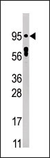
Western blot analysis of anti-Phospho-STAT5 Pab in HepG2 cell line lysate (35ug/lane). Phospho-STAT5(arrow) was detected using the purified Pab (1:60 dilution).Application
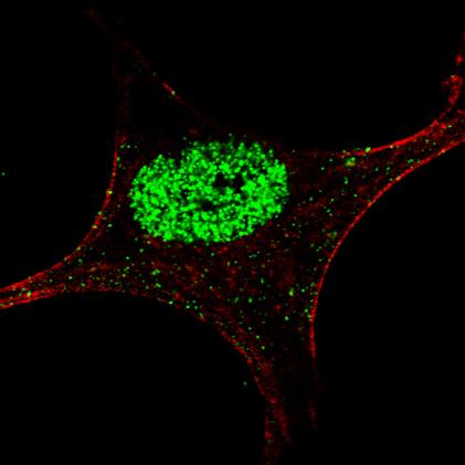
Fluorescent confocal image of an SY5Y cell stained with AP3268a Phospho-STAT5a antibody. SY5Y cells were fixed with 4% PFA (20 min), permeabilized with Triton X-100 (0.2%, 30 min), then incubated with AP3268a Phospho-STAT5a primary antibody (1:200, 2 h at room temperature). For secondary antibody, Alexa Fluor® 488 conjugated donkey anti-rabbit antibody (green) was used (1:1000, 1h).Cytoplasmic actin was counterstained with Alexa Fluor® 555 (red) conjugated Phalloidin (5.25 μM, 25 min). Nuclei were counterstained with Hoechst 33342 (blue) (10 µg/ml, 3 min)(not shown). Phospho-STAT5a immunoreactivity is localized exclusively to the nucleus.Application
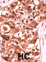
Formalin-fixed and paraffin-embedded human cancer tissue reacted with the primary antibody, which was peroxidase-conjugated to the secondary antibody, followed by DAB staining. This data demonstrates the use of this antibody for immunohistochemistry; clinical relevance has not been evaluated. BC = breast carcinoma; HC = hepatocarcinoma.| Product Name | Phospho-STAT5a-Y694 Antibody |
|---|---|
| Antibody Type | Primary Antibodies |
| Antigen Alias | STAT5A; STAT5; Signal transducer and activator of transcription 5A |
| Modification | p-Y694 |
| Clonality | Polyclonal |
|---|---|
| Isotype | Ig |
| Host Species | Rabbit |
| Tested Applications | WBIFIHC |
| WB:1:100~500 IF |
|
| Species Reactivity | Human |
| Concentration | 1mg/ml |
| Gene Synonyms | STAT5 |
|---|---|
| Alternative Names | STAT5A STAT5 Signal transducer and activator of transcription 5A |
| Molecular Weight(MW) | 90647 Da |
| Function | Carries out a dual function: signal transduction and activation of transcription. Mediates cellular responses to the cytokine KITLG/SCF and other growth factors. Mediates cellular responses to ERBB4. May mediate cellular responses to activated FGFR1, FGFR2, FGFR3 and FGFR4. Binds to the GAS element and activates PRL-induced transcription. Regulates the expression of milk proteins during lactation |
| Tissue Specificity | This Phospho-STAT5a-Y694 antibody is generated from rabbits immunized with a KLH conjugated synthetic phosphopeptide corresponding to amino acid residues surrounding Y694 of human STAT5a. |
| Cellular Localization | Cytoplasm. Nucleus. Note=Translocated into the nucleus in response to phosphorylation |
| Entrez Gene | 6776 |
|---|
Application
Western blot analysis of anti-Phospho-STAT5 Pab in HepG2 cell line lysate (35ug/lane). Phospho-STAT5(arrow) was detected using the purified Pab (1:60 dilution).
Application
Fluorescent confocal image of an SY5Y cell stained with AP3268a Phospho-STAT5a antibody. SY5Y cells were fixed with 4% PFA (20 min), permeabilized with Triton X-100 (0.2%, 30 min), then incubated with AP3268a Phospho-STAT5a primary antibody (1:200, 2 h at room temperature). For secondary antibody, Alexa Fluor® 488 conjugated donkey anti-rabbit antibody (green) was used (1:1000, 1h).Cytoplasmic actin was counterstained with Alexa Fluor® 555 (red) conjugated Phalloidin (5.25 μM, 25 min). Nuclei were counterstained with Hoechst 33342 (blue) (10 µg/ml, 3 min)(not shown). Phospho-STAT5a immunoreactivity is localized exclusively to the nucleus.
Application
Formalin-fixed and paraffin-embedded human cancer tissue reacted with the primary antibody, which was peroxidase-conjugated to the secondary antibody, followed by DAB staining. This data demonstrates the use of this antibody for immunohistochemistry; clinical relevance has not been evaluated. BC = breast carcinoma; HC = hepatocarcinoma.| Application Notes | WB:1:100~500 IF |
|---|
| Form | Liquid |
|---|---|
| Storage Instructions | For short-term storage, store at 4° C. For long-term storage, aliquot and store at -20ºC or below. Avoid multiple freeze-thaw cycles. |
| Storage Buffer | Purified polyclonal antibody supplied in PBS with 0.09% (W/V) sodium azide. This antibody is first purified by protein G affinity chromatography. Then, the antibody fraction is peptide affinity purified in a 2-step procedure with control and phosphorylated peptides. The phospho-specific antibody is eluted with high and low pH buffers and neutralized immediately, followed by dialysis against PBS. |


