Application
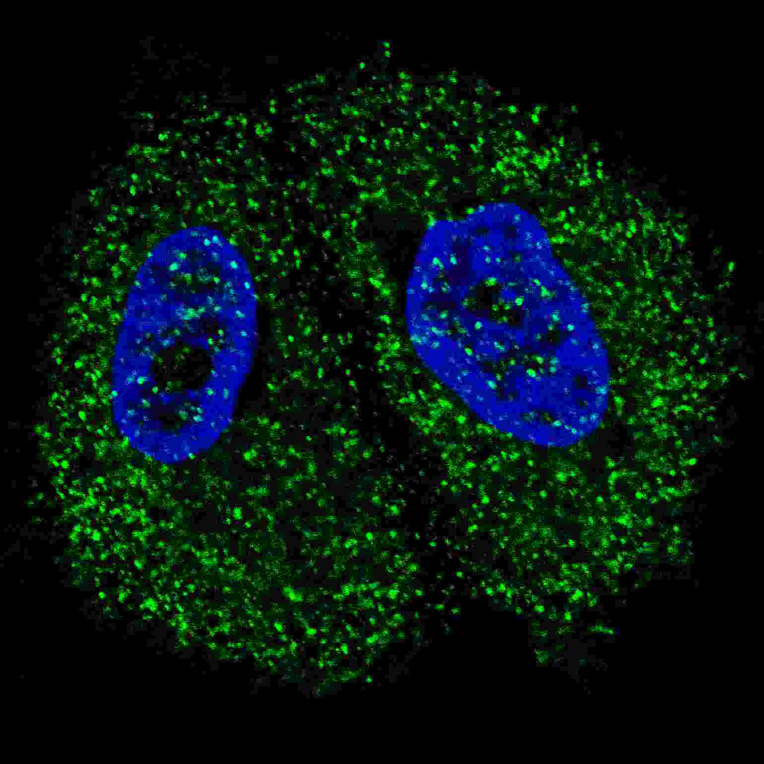
Fluorescent confocal image of MCF7 cells stained with Pyruvate Kinase (PKM2) (C-term) antibody. MCF7 cells were fixed with 4% PFA (20 min), permeabilized with Triton X-100 (0.2%, 30 min). Cells were then incubated with AP7044b Pyruvate Kinase (PKM2) (C-term) primary antibody (1:200, 2 h at room temperature). For secondary antibody, Alexa Fluor® 488 conjugated donkey anti-rabbit antibody (green) was used (1:1000, 1h). Nuclei were counterstained with Hoechst 33342 (blue) (10 μg/ml, 5 min). Note the highly specific localization of the Pyruvate Kinase (PKM2) mainly to the cytoplasm, supported by Human Protein Atlas Data (http://www.proteinatlas.org/ENSG00000067225).Application
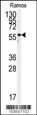
Western blot analysis of anti-h PKM2-N491 Pab in Ramos cell line lysates (35ug/lane). hPKM2-N491(arrow) was detected using the purified Pab (1:60dilution).Application
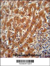
Pyruvate Kinase (PKM2) Antibody (C-term) immunohistochemistry analysis in formalin fixed and paraffin embedded human hepatocarcinoma followed by peroxidase conjugation of the secondary antibody and DAB staining. This data demonstrates the use of the Pyruvate Kinase (PKM2) Antibody (C-term) for immunohistochemistry. Clinical relevance has not been evaluated.| Product Name | Pyruvate Kinase (PKM2) Antibody (C-term) |
|---|---|
| Antibody Type | Primary Antibodies |
| Clonality | Polyclonal |
|---|---|
| Isotype | Ig |
| Host Species | Rabbit |
| Tested Applications | IFWBIHC |
| IF:1:200 WB |
|
| Species Reactivity | Human |
| Concentration | 1mg/ml |
| Gene Synonyms | OIP3, PK2, PK3, PKM2 |
|---|---|
| Alternative Names | PKM OIP3 PK2 PK3 PKM2 Pyruvate kinase isozymes M1/M2 Cytosolic thyroid hormone-binding protein Opa-interacting protein 3 Pyruvate kinase 2/3 Pyruvate kinase muscle isozyme Thyroid hormone-binding protein 1 Tumor M2-PK p58 |
| Molecular Weight(MW) | 57937 Da |
| Function | Glycolytic enzyme that catalyzes the transfer of a phosphoryl group from phosphoenolpyruvate (PEP) to ADP, generating ATP. Stimulates POU5F1-mediated transcriptional activation. Plays a general role in caspase independent cell death of tumor cells The ratio betwween the highly active tetrameric form and nearly inactive dimeric form determines whether glucose carbons are channeled to biosynthetic processes or used for glycolytic ATP production. The transition between the 2 forms contributes to the control of glycolysis and is important for tumor cell proliferation and survival |
| Tissue Specificity | Specifically expressed in proliferating cells, such as embryonic stem cells, embryonic carcinoma cells, as well as cancer cells |
| Cellular Localization | Cytoplasm. Nucleus. Note=Translocates to the nucleus in response to different apoptotic stimuli. Nuclear translocation is sufficient to induce cell death that is caspase independent, isoform-specific and independent of its enzymatic activity |
Application
Fluorescent confocal image of MCF7 cells stained with Pyruvate Kinase (PKM2) (C-term) antibody. MCF7 cells were fixed with 4% PFA (20 min), permeabilized with Triton X-100 (0.2%, 30 min). Cells were then incubated with AP7044b Pyruvate Kinase (PKM2) (C-term) primary antibody (1:200, 2 h at room temperature). For secondary antibody, Alexa Fluor® 488 conjugated donkey anti-rabbit antibody (green) was used (1:1000, 1h). Nuclei were counterstained with Hoechst 33342 (blue) (10 μg/ml, 5 min). Note the highly specific localization of the Pyruvate Kinase (PKM2) mainly to the cytoplasm, supported by Human Protein Atlas Data (http://www.proteinatlas.org/ENSG00000067225).
Application
Western blot analysis of anti-h PKM2-N491 Pab in Ramos cell line lysates (35ug/lane). hPKM2-N491(arrow) was detected using the purified Pab (1:60dilution).
Application
Pyruvate Kinase (PKM2) Antibody (C-term) immunohistochemistry analysis in formalin fixed and paraffin embedded human hepatocarcinoma followed by peroxidase conjugation of the secondary antibody and DAB staining. This data demonstrates the use of the Pyruvate Kinase (PKM2) Antibody (C-term) for immunohistochemistry. Clinical relevance has not been evaluated.| Application Notes | IF:1:200 WB |
|---|
| Form | Liquid |
|---|---|
| Storage Instructions | For short-term storage, store at 4° C. For long-term storage, aliquot and store at -20ºC or below. Avoid multiple freeze-thaw cycles. |
| Storage Buffer | Purified polyclonal antibody supplied in PBS with 0.09% (W/V) sodium azide. This antibody is purified through a protein G column, eluted with high and low pH buffers and neutralized immediately, followed by dialysis against PBS. |


