Application
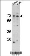
Western blot analysis of hRIPK1-K585 in Jurkat and K562 cell line lysates (35ug/lane). RIPK1 (arrow) was detected using the purified Pab.Application
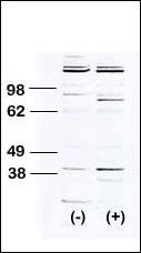
Western blot analysis of anti-RIPK1 Pab transiently transfected HEK-293 cell line lysate (1ug/lane). RIPK1 pab(arrow) was detected using the purified Pab.Application
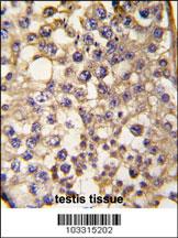
Formalin-fixed and paraffin-embedded human testis tissue reacted with RIPK1 antibody (C-term), which was peroxidase-conjugated to the secondary antibody, followed by DAB staining. This data demonstrates the use of this antibody for immunohistochemistry; clinical relevance has not been evaluated.| Product Name | RIPK1 Antibody (C-term) |
|---|---|
| Antibody Type | Primary Antibodies |
| Clonality | Polyclonal |
|---|---|
| Isotype | Ig |
| Host Species | Rabbit |
| Tested Applications | WBIHC |
| WB:1:250~500 IHC |
|
| Species Reactivity | Human |
| Concentration | 1mg/ml |
| Gene Synonyms | RIP, RIP1 |
|---|---|
| Alternative Names | RIPK1 RIP RIP1 Receptor-interacting serine/threonine-protein kinase 1 Cell death protein RIP Receptor-interacting protein 1 Serine/threonine-protein kinase RIP |
| Molecular Weight(MW) | 75931 Da |
| Function | Serine-threonine kinase which transduces inflammatory and cell-death signals (programmed necrosis) following death receptors ligation, activation of pathogen recognition receptors (PRRs), and DNA damage. Upon activation of TNFR1 by the TNF-alpha family cytokines, TRADD and TRAF2 are recruited to the receptor Ubiquitination by TRAF2 via 'Lys-63'-link chains acts as a critical enhancer of communication with downstream signal transducers in the mitogen-activated protein kinase pathway and the NF-kappa-B pathway, which in turn mediate downstream events including the activation of genes encoding inflammatory molecules Polyubiquitinated protein binds to IKBKG/NEMO, the regulatory subunit of the IKK complex, a critical event for NF-kappa-B activation. Interaction with other cellular RHIM-containing adapters initiates gene activation and cell death. RIPK1 and RIPK3 association, in particular, forms a necrosis-inducing complex |
| Tissue Specificity | This RIPK1 antibody is generated from rabbits immunized with a KLH conjugated synthetic peptide between 585~671 amino acids from the C-terminal region of human RIPK1. |
| Cellular Localization | Cytoplasm. Cell membrane (By similarity). |
Application
Western blot analysis of hRIPK1-K585 in Jurkat and K562 cell line lysates (35ug/lane). RIPK1 (arrow) was detected using the purified Pab.
Application
Western blot analysis of anti-RIPK1 Pab transiently transfected HEK-293 cell line lysate (1ug/lane). RIPK1 pab(arrow) was detected using the purified Pab.
Application
Formalin-fixed and paraffin-embedded human testis tissue reacted with RIPK1 antibody (C-term), which was peroxidase-conjugated to the secondary antibody, followed by DAB staining. This data demonstrates the use of this antibody for immunohistochemistry; clinical relevance has not been evaluated.| Application Notes | WB:1:250~500 IHC |
|---|
| Form | Liquid |
|---|---|
| Storage Instructions | For short-term storage, store at 4° C. For long-term storage, aliquot and store at -20ºC or below. Avoid multiple freeze-thaw cycles. |
| Storage Buffer | Purified polyclonal antibody supplied in PBS with 0.09% (W/V) sodium azide. This antibody is purified through a protein G column, eluted with high and low pH buffers and neutralized immediately, followed by dialysis against PBS. |


