Application
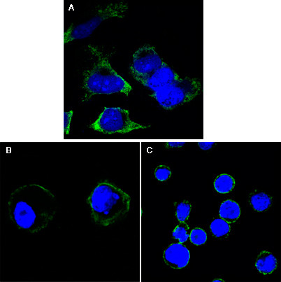
Figure 1: Confocal immunofluorescence analysis of Hela (A), A431 (B) and THP-1 (C) cells using anti-RTN3 monoclonal antibody (green). Blue: DRAQ5 fluorescent DNA dye.| Product Name | RTN3 Antibody |
|---|---|
| Antibody Type | Primary Antibodies |
| Antigen Alias | HAP; ASYIP; NSPL2; NSPLII; RTN3-A1 |
| Clonality | Monoclonal |
|---|---|
| Isotype | IgG2a |
| Clone Number | 100000000000 |
| Host Species | Mouse |
| Tested Applications | IF |
| IF:1:200 - 1000. |
|
| Species Reactivity | Human |
| Concentration | 1mg/ml |
| Gene Synonyms | ASYIP NSPL2 |
|---|---|
| Alternative Names | HAP ASYIP NSPL2 NSPLII RTN3-A1 |
| Molecular Weight(MW) | 112611 Da |
| Function | May be involved in membrane trafficking in the early secretory pathway. Inhibits BACE1 activity and amyloid precursor protein processing. May induce caspase-8 cascade and apoptosis May favor BCL2 translocation to the mitochondria upon endoplasmic reticulum stress. In case of enteroviruses infection, RTN3 may be involved in the viral replication or pathogenesis |
| Tissue Specificity | Isoform 3 is widely expressed, with highest levels in brain, where it is enriched in neuronal cell bodies from gray matter (at protein level). Three times more abundant in macula than in peripheral retina. Isoform 1 is expressed at high levels in brain and at low levels in skeletal muscle. Isoform 2 is only found in melanoma |
| Cellular Localization | Endoplasmic reticulum membrane; Multi-pass membrane protein. Golgi apparatus membrane; Multi-pass membrane protein |
| Entrez Gene | 10313 |
|---|---|
| Nucleotide Accession | NP_958833.1 |
Application
Figure 1: Confocal immunofluorescence analysis of Hela (A), A431 (B) and THP-1 (C) cells using anti-RTN3 monoclonal antibody (green). Blue: DRAQ5 fluorescent DNA dye.| Application Notes | IF:1:200 - 1000. |
|---|
| Form | Liquid |
|---|---|
| Storage Instructions | For short-term storage, store at 4° C. For long-term storage, aliquot and store at -20ºC or below. Avoid multiple freeze-thaw cycles. |
| Storage Buffer | Purified recombinant fragment of RTN3 expressed in E. Coli. |


