Application
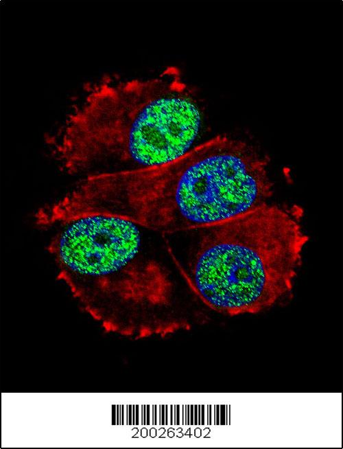
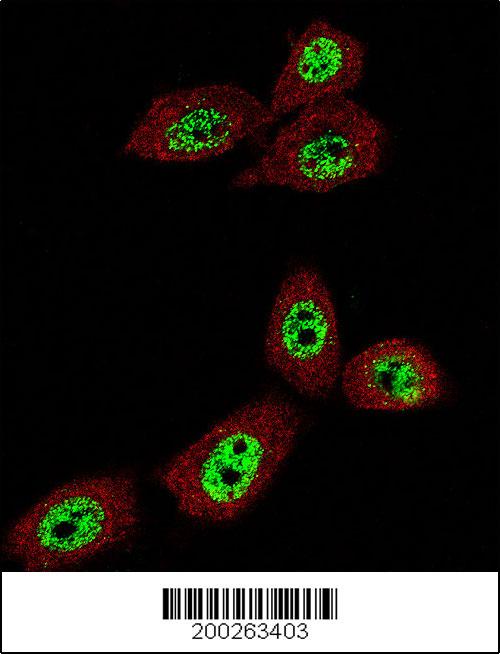
Fluorescent confocal image of SY5Y cells stained with SOX2 antibody. SY5Y cells were fixed with 4% PFA (20 min), permeabilized with Triton X-100 (0.2%, 30 min). Cells were then incubated with AM2048a SOX2 primary antibody (1:100, 2 h at room temperature). For secondary antibody, Alexa Fluor® 488 conjugated donkey anti-mouse antibody (green) was used (1:1000, 1h). Note the highly specific localization of the SOX2 mainly to the nucleus.Application
Confocal immunofluorescent analysis of SOX2 Antibody(Cat#AM2048a) with MCF-7 cell followed by Alexa Fluor® 488-conjugated goat anti-mouse lgG (green).Actin filaments have been labeled with Alexa Fluor® 555 phalloidin (red). DAPI was used to stain the cell nuclear (blue).Application
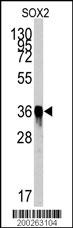
Western blot analysis of SOX2 Antibody (Cat.#AM2048a) by SOX2 recombinant protein. SOX2(arrow) was detected using the purified Mab.Application
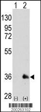
Western blot analysis of SOX2 (arrow) using mouse monoclonal SOX2 antibody (Cat.#AM2048a). 293 cell lysates (2 ug/lane) either nontransfected (Lane 1) or transiently transfected with the SOX2 gene (Lane 2) (Origene Technologies)Application
Confocal immunofluorescent analysis of SOX2 Antibody(Cat#AM2048a) with U-251MG cell followed by Alexa Fluor® 488-conjugated goat anti-mouse lgG (green).Actin filaments have been labeled with Alexa Fluor® 555 phalloidin (red).Application
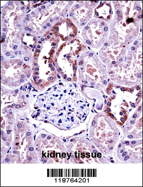
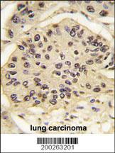
Formalin-fixed and paraffin-embedded human lung carcinoma tissue reacted with SOX2 Antibody (Cat.#AM2048a), which was peroxidase-conjugated to the secondary antibody, followed by DAB staining. This data demonstrates the use of this antibody for immunohistochemistry; clinical relevance has not been evaluated.Application
Flow cytometric analysis of NCI-H460 cells using SOX2 Monoclonal Antibody (bottom histogram) compared to a negative control cell (top histogram). PE-conjugated goat-anti-mouse secondary antibodies were used for the analysis.| Product Name | SOX2 Antibody |
|---|---|
| Antibody Type | Primary Antibodies |
| Antigen Alias | SOX2; Transcription factor SOX-2 |
| Clonality | Monoclonal |
|---|---|
| Isotype | IgG1 |
| Clone Number | 57CT23.3.4 |
| Host Species | Mouse |
| Tested Applications | IFWBIHCFC |
| WB:1:500~1:1000 IF:1:100~1:500 IHC:1:50~1:100 |
|
| Species Reactivity | Human |
| Concentration | 1mg/ml |
| Alternative Names | SOX2 Transcription factor SOX-2 |
|---|---|
| Molecular Weight(MW) | 34310 Da |
| Function | Transcription factor that forms a trimeric complex with OCT4 on DNA and controls the expression of a number of genes involved in embryonic development such as YES1, FGF4, UTF1 and ZFP206 (By similarity). Critical for early embryogenesis and for embryonic stem cell pluripotency. May function as a switch in neuronal development. Downstream SRRT target that mediates the promotion of neural stem cell self-renewal (By similarity). Keeps neural cells undifferentiated by counteracting the activity of proneural proteins and suppresses neuronal differentiation (By similarity) |
| Tissue Specificity | SOX2 recombinant protein is used to produce this monoclonal antibody. |
| Cellular Localization | Nucleus. |
| Entrez Gene | 6657 |
|---|
Application
Fluorescent confocal image of SY5Y cells stained with SOX2 antibody. SY5Y cells were fixed with 4% PFA (20 min), permeabilized with Triton X-100 (0.2%, 30 min). Cells were then incubated with AM2048a SOX2 primary antibody (1:100, 2 h at room temperature). For secondary antibody, Alexa Fluor® 488 conjugated donkey anti-mouse antibody (green) was used (1:1000, 1h). Note the highly specific localization of the SOX2 mainly to the nucleus.
Application
Confocal immunofluorescent analysis of SOX2 Antibody(Cat#AM2048a) with MCF-7 cell followed by Alexa Fluor® 488-conjugated goat anti-mouse lgG (green).Actin filaments have been labeled with Alexa Fluor® 555 phalloidin (red). DAPI was used to stain the cell nuclear (blue).
Application
Western blot analysis of SOX2 Antibody (Cat.#AM2048a) by SOX2 recombinant protein. SOX2(arrow) was detected using the purified Mab.
Application
Western blot analysis of SOX2 (arrow) using mouse monoclonal SOX2 antibody (Cat.#AM2048a). 293 cell lysates (2 ug/lane) either nontransfected (Lane 1) or transiently transfected with the SOX2 gene (Lane 2) (Origene Technologies)
Application
Confocal immunofluorescent analysis of SOX2 Antibody(Cat#AM2048a) with U-251MG cell followed by Alexa Fluor® 488-conjugated goat anti-mouse lgG (green).Actin filaments have been labeled with Alexa Fluor® 555 phalloidin (red).
Application
Formalin-fixed and paraffin-embedded human lung carcinoma tissue reacted with SOX2 Antibody (Cat.#AM2048a), which was peroxidase-conjugated to the secondary antibody, followed by DAB staining. This data demonstrates the use of this antibody for immunohistochemistry; clinical relevance has not been evaluated.
Application
Flow cytometric analysis of NCI-H460 cells using SOX2 Monoclonal Antibody (bottom histogram) compared to a negative control cell (top histogram). PE-conjugated goat-anti-mouse secondary antibodies were used for the analysis.| Application Notes | WB:1:500~1:1000 IF:1:100~1:500 IHC:1:50~1:100 |
|---|
| Form | Liquid |
|---|---|
| Storage Instructions | For short-term storage, store at 4° C. For long-term storage, aliquot and store at -20ºC or below. Avoid multiple freeze-thaw cycles. |
| Storage Buffer | Purified monoclonal antibody supplied in PBS with 0.09% (W/V) sodium azide. This antibody is purified through a protein G column, eluted with high and low pH buffers and neutralized immediately, followed by dialysis against PBS. |


