Application
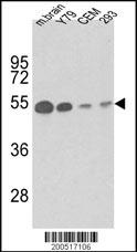
Western blot analysis of anti-TBB1 Antibody (Cat. #AM1013a) in mouse brain tissue, Y79, CEM and 293 lysates (35ug/lane). TBB1 (arrow) was detected using the purified Mab.(1ug/ml)Application
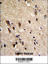
Formalin-fixed and paraffin-embedded human brain reacted with TBB1 Antibody, which was peroxidase-conjugated to the secondary antibody, followed by DAB staining. This data demonstrates the use of this antibody for immunohistochemistry; clinical relevance has not been evaluated.Application
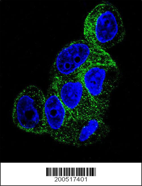
Confocal immunofluorescent analysis of TBB1 Antibody (Cat#AM1013a) with HepG2 cell followed by Alexa Fluor® 488-conjugated goat anti-mouse lgG (green). DAPI was used to stain the cell nuclear (blue).| Product Name | TBB1 Antibody |
|---|---|
| Antibody Type | Primary Antibodies |
| Antigen Alias | TUBB1; Tubulin beta-1 chain |
| Clonality | Monoclonal |
|---|---|
| Isotype | IgG1 |
| Clone Number | 86CT4.4.4 |
| Host Species | Mouse |
| Tested Applications | WBIHCIF |
| WB:1:100~500 IHC |
|
| Species Reactivity | Human |
| Concentration | 1mg/ml |
| Alternative Names | TUBB1 Tubulin beta-1 chain |
|---|---|
| Molecular Weight(MW) | 50327 Da |
| Function | Tubulin is the major constituent of microtubules. It binds two moles of GTP, one at an exchangeable site on the beta chain and one at a non-exchangeable site on the alpha chain (By similarity) |
| Tissue Specificity | Hematopoietic cell-specific. Major isotype in leukocytes, where it represents 50% of all beta-tubulins |
| Cellular Localization | Cytoplasm, cytoskeleton. |
| Entrez Gene | 81027 |
|---|
Application
Western blot analysis of anti-TBB1 Antibody (Cat. #AM1013a) in mouse brain tissue, Y79, CEM and 293 lysates (35ug/lane). TBB1 (arrow) was detected using the purified Mab.(1ug/ml)
Application
Formalin-fixed and paraffin-embedded human brain reacted with TBB1 Antibody, which was peroxidase-conjugated to the secondary antibody, followed by DAB staining. This data demonstrates the use of this antibody for immunohistochemistry; clinical relevance has not been evaluated.
Application
Confocal immunofluorescent analysis of TBB1 Antibody (Cat#AM1013a) with HepG2 cell followed by Alexa Fluor® 488-conjugated goat anti-mouse lgG (green). DAPI was used to stain the cell nuclear (blue).| Application Notes | WB:1:100~500 IHC |
|---|
| Form | Liquid |
|---|---|
| Storage Instructions | For short-term storage, store at 4° C. For long-term storage, aliquot and store at -20ºC or below. Avoid multiple freeze-thaw cycles. |
| Storage Buffer | Purified monoclonal antibody supplied in PBS with 0.09% (W/V) sodium azide. This antibody is purified through a protein G column, eluted with high and low pH buffers and neutralized immediately, followed by dialysis against PBS. |


