Application
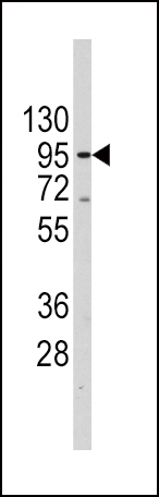
Western blot analysis of anti-TERT Antibody (Center) (RB14069) in Jurkat cell line lysates (35ug/lane). TERT(arrow) was detected using the purified Pab (1:60 dilution).Application
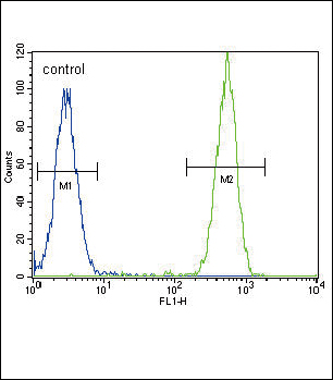
TERT Antibody (Center) flow cytometric analysis of Hela cells (right histogram) compared to a negative control cell (left histogram).FITC-conjugated goat-anti-rabbit secondary antibodies were used for the analysis.Application
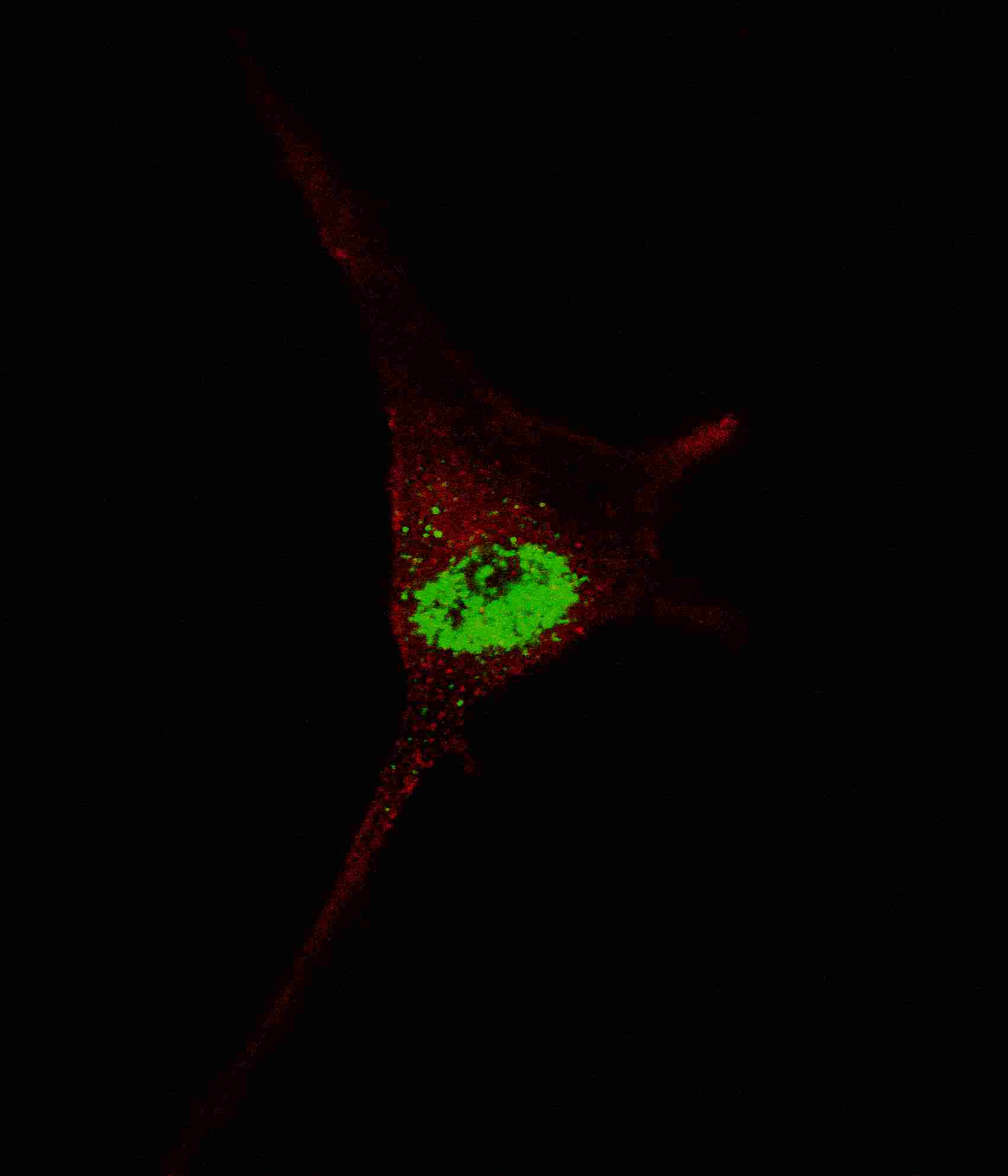
Fluorescent confocal image of SY5Y cells stained with TERT (Center) antibody. SY5Y cells were fixed with 4% PFA (20 min), permeabilized with Triton X-100 (0.2%, 30 min). Cells were then incubated with AP1410c TERT (Center) primary antibody (1:200, 2 h at room temperature). For secondary antibody, Alexa Fluor® 488 conjugated donkey anti-rabbit antibody (green) was used (1:1000, 1h). Nuclei were counterstained with Hoechst 33342 (blue) (10 μg/ml, 5 min). TERT immunoreactivity is localized to the nucleus of SY5Y cells.Application
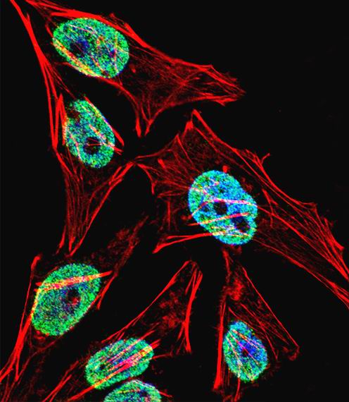
Fluorescent confocal image of Hela cell stained with TERT Antibody (Center)(Cat#AP1410c).Hela cells were fixed with 4% PFA (20 min), permeabilized with Triton X-100 (0.1%, 10 min), then incubated with TERT primary antibody (1:25, 1 h at 37℃). For secondary antibody, Alexa Fluor® 488 conjugated donkey anti-rabbit antibody (green) was used (1:400, 50 min at 37℃).Cytoplasmic actin was counterstained with Alexa Fluor® 555 (red) conjugated Phalloidin (7units/ml, 1 h at 37℃). Nuclei were counterstained with DAPI (blue) (10 µg/ml, 10 min). TERT immunoreactivity is localized to Nucleus significantly and Cytoplasm weakly.| Product Name | TERT Antibody (Center) |
|---|---|
| Antibody Type | Primary Antibodies |
| Clonality | Polyclonal |
|---|---|
| Isotype | Ig |
| Host Species | Rabbit |
| Tested Applications | WBFCIF |
| WB:1:100~500 FC |
|
| Species Reactivity | Human |
| Concentration | 1mg/ml |
| Gene Synonyms | EST2, TCS1, TRT |
|---|---|
| Alternative Names | TERT EST2 TCS1 TRT Telomerase reverse transcriptase HEST2 Telomerase catalytic subunit Telomerase-associated protein 2 |
| Molecular Weight(MW) | 126997 Da |
| Function | Telomerase is a ribonucleoprotein enzyme essential for the replication of chromosome termini in most eukaryotes. Active in progenitor and cancer cells. Inactive, or very low activity, in normal somatic cells. Catalytic component of the teleromerase holoenzyme complex whose main activity is the elongation of telomeres by acting as a reverse transcriptase that adds simple sequence repeats to chromosome ends by copying a template sequence within the RNA component of the enzyme. Catalyzes the RNA- dependent extension of 3'-chromosomal termini with the 6- nucleotide telomeric repeat unit, 5'-TTAGGG-3'. The catalytic cycle involves primer binding, primer extension and release of product once the template boundary has been reached or nascent product translocation followed by further extension. More active on substrates containing 2 or 3 telomeric repeats. Telomerase activity is regulated by a number of factors including telomerase complex-associated proteins, chaperones and polypeptide modifiers Modulates Wnt signaling. Plays important roles in aging and antiapoptosis |
| Tissue Specificity | Expressed at a high level in thymocyte subpopulations, at an intermediate level in tonsil T-lymphocytes, and at a low to undetectable level in peripheral blood T- lymphocytes |
| Cellular Localization | Nucleus, nucleolus. Nucleus, nucleoplasm. Nucleus. Chromosome, telomere. Cytoplasm. Nucleus, PML body Note=Shuttling between nuclear and cytoplasm depends on cell cycle, phosphorylation states, transformation and DNA damage Diffuse localization in the nucleoplasm. Enriched in nucleoli of certain cell types. Translocated to the cytoplasm via nuclear pores in a CRM1/RAN-dependent manner involving oxidative stress- mediated phosphorylation at Tyr-707. Dephosphorylation at this site by SHP2 retains TERT in the nucleus. Translocated to the nucleus by phosphorylation by AKT |
| Entrez Gene | 7015 |
|---|
Application
Western blot analysis of anti-TERT Antibody (Center) (RB14069) in Jurkat cell line lysates (35ug/lane). TERT(arrow) was detected using the purified Pab (1:60 dilution).
Application
TERT Antibody (Center) flow cytometric analysis of Hela cells (right histogram) compared to a negative control cell (left histogram).FITC-conjugated goat-anti-rabbit secondary antibodies were used for the analysis.
Application
Fluorescent confocal image of SY5Y cells stained with TERT (Center) antibody. SY5Y cells were fixed with 4% PFA (20 min), permeabilized with Triton X-100 (0.2%, 30 min). Cells were then incubated with AP1410c TERT (Center) primary antibody (1:200, 2 h at room temperature). For secondary antibody, Alexa Fluor® 488 conjugated donkey anti-rabbit antibody (green) was used (1:1000, 1h). Nuclei were counterstained with Hoechst 33342 (blue) (10 μg/ml, 5 min). TERT immunoreactivity is localized to the nucleus of SY5Y cells.
Application
Fluorescent confocal image of Hela cell stained with TERT Antibody (Center)(Cat#AP1410c).Hela cells were fixed with 4% PFA (20 min), permeabilized with Triton X-100 (0.1%, 10 min), then incubated with TERT primary antibody (1:25, 1 h at 37℃). For secondary antibody, Alexa Fluor® 488 conjugated donkey anti-rabbit antibody (green) was used (1:400, 50 min at 37℃).Cytoplasmic actin was counterstained with Alexa Fluor® 555 (red) conjugated Phalloidin (7units/ml, 1 h at 37℃). Nuclei were counterstained with DAPI (blue) (10 µg/ml, 10 min). TERT immunoreactivity is localized to Nucleus significantly and Cytoplasm weakly.| Application Notes | WB:1:100~500 FC |
|---|
| Form | Liquid |
|---|---|
| Storage Instructions | For short-term storage, store at 4° C. For long-term storage, aliquot and store at -20ºC or below. Avoid multiple freeze-thaw cycles. |
| Storage Buffer | Purified polyclonal antibody supplied in PBS with 0.09% (W/V) sodium azide. This antibody is prepared by Saturated Ammonium Sulfate (SAS) precipitation followed by dialysis against PBS. |


