Application
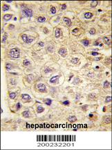
Formalin-fixed and paraffin-embedded human hepatocarcinoma tissue reacted with TYRO3 Antibody (Cat.#AM7679b), which was peroxidase-conjugated to the secondary antibody, followed by DAB staining. This data demonstrates the use of this antibody for immunohistochemistry; clinical relevance has not been evaluated.Application
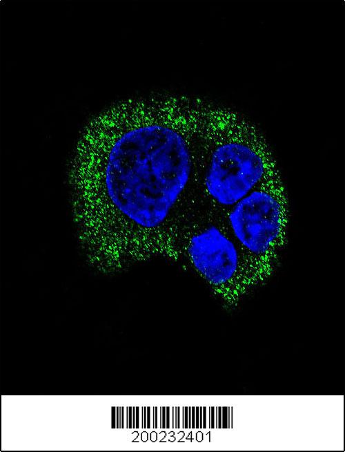
Confocal immunofluorescent analysis of TYRO3 Antibody (Cat#AM7679b) with HepG2 cell followed by Alexa Fluor 488-conjugated goat anti-mouse lgG (green). DAPI was used to stain the cell nuclear (blue).IHC
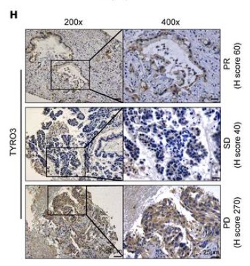
(H) IHC staining of TYRO3 in patients with lung cancer who received anti–PD-1/PD-L1 therapy. H score for TYRO3 expression in patients who were resistant or nonresistant to anti–PD-1/PD-L1 therapy. n = 12 resistant patients; n = 17 nonresistant patients. *P = 0.0132, by 2-tailed, unpaired Student’s t test. Scale bars: 50 μm (left) and 25 μm (right). Data are presented as the mean ± SD.| Product Name | TYRO3 Antibody |
|---|---|
| Antibody Type | Primary Antibodies |
| Clonality | Monoclonal |
|---|---|
| Clone Number | 64CT15.6.2.3.1 |
| Host Species | Mouse |
| Tested Applications | IFIHC |
| IHC:1:50~100 IF |
|
| Species Reactivity | Human |
| Concentration | 1mg/ml |
| Gene Synonyms | BYK DTK RSE SKY TIF |
|---|---|
| Alternative Names | TYRO3 SKY BYK DTK RSE Tyrosine-protein kinase receptor TYRO3 Tyrosine-protein kinase DTK Tyrosine-protein kinase RSE Tyrosine-protein kinase SKY Tyrosine-protein kinase byk |
| Molecular Weight(MW) | 96905 Da |
| Function | Receptor tyrosine kinase that transduces signals from the extracellular matrix into the cytoplasm by binding to several ligands including TULP1 or GAS6. Regulates many physiological processes including cell survival, migration and differentiation Ligand binding at the cell surface induces dimerization and autophosphorylation of TYRO3 on its intracellular domain that provides docking sites for downstream signaling molecules Following activation by ligand, interacts with PIK3R1 and thereby enhances PI3-kinase activity. Activates the AKT survival pathway, including nuclear translocation of NF-kappa-B and up-regulation of transcription of NF-kappa-B-regulated genes. TYRO3 signaling plays a role in various processes such as neuron protection from excitotoxic injury, platelet aggregation and cytoskeleton reorganization. Plays also an important role in inhibition of Toll-like receptors (TLRs)-mediated innate immune response by activating STAT1, which selectively induces production of suppressors of cytokine signaling SOCS1 and SOCS3 |
| Tissue Specificity | Abundant in the brain and lower levels in other tissues |
| Cellular Localization | Cell membrane; Single-pass type I membrane protein |
Application
Formalin-fixed and paraffin-embedded human hepatocarcinoma tissue reacted with TYRO3 Antibody (Cat.#AM7679b), which was peroxidase-conjugated to the secondary antibody, followed by DAB staining. This data demonstrates the use of this antibody for immunohistochemistry; clinical relevance has not been evaluated.
Application
Confocal immunofluorescent analysis of TYRO3 Antibody (Cat#AM7679b) with HepG2 cell followed by Alexa Fluor 488-conjugated goat anti-mouse lgG (green). DAPI was used to stain the cell nuclear (blue).
IHC
(H) IHC staining of TYRO3 in patients with lung cancer who received anti–PD-1/PD-L1 therapy. H score for TYRO3 expression in patients who were resistant or nonresistant to anti–PD-1/PD-L1 therapy. n = 12 resistant patients; n = 17 nonresistant patients. *P = 0.0132, by 2-tailed, unpaired Student’s t test. Scale bars: 50 μm (left) and 25 μm (right). Data are presented as the mean ± SD.| Application Notes | IHC:1:50~100 IF |
|---|
| Form | Liquid |
|---|---|
| Storage Instructions | For short-term storage, store at 4° C. For long-term storage, aliquot and store at -20ºC or below. Avoid multiple freeze-thaw cycles. |
| Storage Buffer | Purified mouse monoclonal antibody supplied in PBS with 0.09% (W/V) sodium azide. This antibody is prepared by Saturated Ammonium Sulfate (SAS) precipitation followed by dialysis against PBS. |


