Application
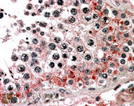
Western blot analysis of anti-VGFR3 Monoclonal Antibody by VGFR3 recombinant protein(Fragment 50KD). VGFR3 protein (arrow) was detected using the ascites Mab.| Product Name | VGFR3 Antibody (Ascites) |
|---|---|
| Antibody Type | Primary Antibodies |
| Clonality | Monoclonal |
|---|---|
| Isotype | IgG1 |
| Clone Number | 44CT92.4.4 |
| Host Species | Mouse |
| Tested Applications | IHCWB |
| WB:1:1000 IHC |
|
| Species Reactivity | Human |
| Concentration | 1mg/ml |
| Purification | Affinity purified |
| Gene Synonyms | VEGFR3 |
|---|---|
| Alternative Names | FLT4 VEGFR3 Vascular endothelial growth factor receptor 3 Fms-like tyrosine kinase 4 Tyrosine-protein kinase receptor FLT4 |
| Molecular Weight(MW) | 152757 Da |
| Function | Tyrosine-protein kinase that acts as a cell-surface receptor for VEGFC and VEGFD, and plays an essential role in adult lymphangiogenesis and in the development of the vascular network and the cardiovascular system during embryonic development Promotes proliferation, survival and migration of endothelial cells, and regulates angiogenic sprouting. Signaling by activated FLT4 leads to enhanced production of VEGFC, and to a lesser degree VEGFA, thereby creating a positive feedback loop that enhances FLT4 signaling. Modulates KDR signaling by forming heterodimers The secreted isoform 3 may function as a decoy receptor for VEGFC and/or VEGFD and play an important role as a negative regulator of VEGFC-mediated lymphangiogenesis and angiogenesis. Binding of vascular growth factors to isoform 1 or isoform 2 leads to the activation of several signaling cascades; isoform 2 seems to be less efficient in signal transduction, because it has a truncated C-terminus and therefore lacks several phosphorylation sites Mediates activation of the MAPK1/ERK2, MAPK3/ERK1 signaling pathway, of MAPK8 and the JUN signaling pathway, and of the AKT1 signaling pathway. Phosphorylates SHC1. Mediates phosphorylation of PIK3R1, the regulatory subunit of phosphatidylinositol 3- kinase. Promotes phosphorylation of MAPK8 at 'Thr-183' and 'Tyr- 185', and of AKT1 at 'Ser-473' |
| Tissue Specificity | Detected in endothelial cells (at protein level). Widely expressed. Detected in fetal spleen, lung and brain. Detected in adult liver, muscle, thymus, placenta, lung, testis, ovary, prostate, heart, and kidney |
| Cellular Localization | Cell membrane; Single-pass type I membrane protein. Cytoplasm. Nucleus. Note=Ligand-mediated autophosphorylation leads to rapid internalization Isoform 2: Cell membrane; Single-pass type I membrane protein |
| Entrez Gene | 2324 |
|---|
Application
Western blot analysis of anti-VGFR3 Monoclonal Antibody by VGFR3 recombinant protein(Fragment 50KD). VGFR3 protein (arrow) was detected using the ascites Mab.| Application Notes | WB:1:1000 IHC |
|---|
| Form | Liquid |
|---|---|
| Storage Instructions | For short-term storage, store at 4° C. For long-term storage, aliquot and store at -20ºC or below. Avoid multiple freeze-thaw cycles. |
| Storage Buffer | Mouse monoclonal antibody supplied in crude ascites with 0.09% (W/V) sodium azide. |


