Application
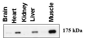
Western blot using anti-AGL (C-term) antibody (AP2402c) at 1:500 dilution. A total of 20 ug of lysates was loaded for each tissue. Data courtesy of Dr. Alan Cheng, Department of Internal Medicine, Life Sciences Institute, University of Michigan Medical Center, Ann Arbor, Michigan.Application
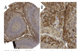
Expression of myc-GS causes wild type but not the ÃCBD mutant of AGL to aggregate around the PAS-stain-positive inclusions. HepG2 cells were transfected with either HA-tagged wild-type AGL (HA-AGL) or HA-AGL ÃCBD. Cells were fixed in formalin and processed for IF using anti-HA (green) and anti-myc (red) antibodies. White arrows indicate colocalization of HA-AGL and myc-GS.Application
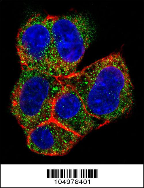
Confocal immunofluorescent analysis of AGL Antibody (C-term)(Cat#AP2402b) with HepG2 cell followed by Alexa Fluor 488-conjugated goat anti-rabbit lgG (green). Actin filaments have been labeled with Alexa Fluor 555 phalloidin (red).DAPI was used to stain the cell nuclear (blue).| Product Name | AGL Antibody (C-term) |
|---|---|
| Antibody Type | Primary Antibodies |
| Clonality | Polyclonal |
|---|---|
| Isotype | Ig |
| Host Species | Rabbit |
| Tested Applications | WBIF |
| WB:1:100-500 IF |
|
| Species Reactivity | Human |
| Concentration | 1mg/ml |
| Gene Synonyms | GDE |
|---|---|
| Alternative Names | AGL GDE Glycogen debranching enzyme Glycogen debrancher 4-alpha-glucanotransferase Oligo-1 4-1 4-glucantransferase Amylo-alpha-1 6-glucosidase Dextrin 6-alpha-D-glucosidase |
| Molecular Weight(MW) | 174764 Da |
| Function | Multifunctional enzyme acting as 1,4-alpha-D-glucan:1,4- alpha-D-glucan 4-alpha-D-glycosyltransferase and amylo-1,6- glucosidase in glycogen degradation |
| Tissue Specificity | Liver, kidney and lymphoblastoid cells express predominantly isoform 1; whereas muscle and heart express not only isoform 1, but also muscle-specific isoform mRNAs (isoforms 2, 3 and 4). Isoforms 5 and 6 are present in both liver and muscle |
| Cellular Localization | Cytoplasm. Note=Under glycogenolytic conditions localizes to the nucleus |
| Entrez Gene | 178 |
|---|
Application
Western blot using anti-AGL (C-term) antibody (AP2402c) at 1:500 dilution. A total of 20 ug of lysates was loaded for each tissue. Data courtesy of Dr. Alan Cheng, Department of Internal Medicine, Life Sciences Institute, University of Michigan Medical Center, Ann Arbor, Michigan.
Application
Expression of myc-GS causes wild type but not the ÃCBD mutant of AGL to aggregate around the PAS-stain-positive inclusions. HepG2 cells were transfected with either HA-tagged wild-type AGL (HA-AGL) or HA-AGL ÃCBD. Cells were fixed in formalin and processed for IF using anti-HA (green) and anti-myc (red) antibodies. White arrows indicate colocalization of HA-AGL and myc-GS.
Application
Confocal immunofluorescent analysis of AGL Antibody (C-term)(Cat#AP2402b) with HepG2 cell followed by Alexa Fluor 488-conjugated goat anti-rabbit lgG (green). Actin filaments have been labeled with Alexa Fluor 555 phalloidin (red).DAPI was used to stain the cell nuclear (blue).| Application Notes | WB:1:100-500 IF |
|---|
| Form | Liquid |
|---|---|
| Storage Instructions | For short-term storage, store at 4° C. For long-term storage, aliquot and store at -20ºC or below. Avoid multiple freeze-thaw cycles. |
| Storage Buffer | Purified polyclonal antibody supplied in PBS with 0.09% (W/V) sodium azide. This antibody is purified through a protein A column, eluted with high and low pH buffers and neutralized immediately, followed by dialysis against PBS. |


