Application
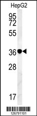
AKR1C1 Antibody (Center) western blot analysis in HepG2 cell line lysates (35ug/lane).This demonstrates the AKR1C1 antibody detected the AKR1C1 protein (arrow).Application
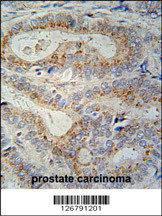
AKR1C1 Antibody (Center) immunohistochemistry analysis in formalin fixed and paraffin embedded human prostate carcinoma followed by peroxidase conjugation of the secondary antibody and DAB staining. This data demonstrates the use of the AKR1C1 Antibody (Center) for immunohistochemistry. Clinical relevance has not been evaluated.Application
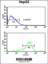
AKR1C1 Antibody (Center) flow cytometric analysis of HepG2 cells (bottom histogram) compared to a negative control cell (top histogram).FITC-conjugated goat-anti-rabbit secondary antibodies were used for the analysis.| Product Name | AKR1C1 Antibody (Center) |
|---|---|
| Antibody Type | Primary Antibodies |
| Clonality | Polyclonal |
|---|---|
| Isotype | Ig |
| Host Species | Rabbit |
| Tested Applications | WBIHCFC |
| WB:1:100~500 IHC |
|
| Species Reactivity | Human |
| Concentration | 1mg/ml |
| Gene Synonyms | DDH, DDH1 |
|---|---|
| Alternative Names | AKR1C1 DDH DDH1 Aldo-keto reductase family 1 member C1 20-alpha-hydroxysteroid dehydrogenase Chlordecone reductase homolog HAKRC Dihydrodiol dehydrogenase 1/2 High-affinity hepatic bile acid-binding protein Indanol dehydrogenase Trans-1 2-dihydro |
| Molecular Weight(MW) | 36788 Da |
| Function | Converts progesterone to its inactive form, 20-alpha- dihydroxyprogesterone (20-alpha-OHP). In the liver and intestine, may have a role in the transport of bile. May have a role in monitoring the intrahepatic bile acid concentration. Has a low bile-binding ability. May play a role in myelin formation |
| Tissue Specificity | Expressed in all tissues tested including liver, prostate, testis, adrenal gland, brain, uterus, mammary gland and keratinocytes. Highest levels found in liver, mammary gland and brain |
| Cellular Localization | Cytoplasm. |
| Entrez Gene | 1645 |
|---|---|
| Nucleotide Accession | NP_001344.2 |
Application
AKR1C1 Antibody (Center) western blot analysis in HepG2 cell line lysates (35ug/lane).This demonstrates the AKR1C1 antibody detected the AKR1C1 protein (arrow).
Application
AKR1C1 Antibody (Center) immunohistochemistry analysis in formalin fixed and paraffin embedded human prostate carcinoma followed by peroxidase conjugation of the secondary antibody and DAB staining. This data demonstrates the use of the AKR1C1 Antibody (Center) for immunohistochemistry. Clinical relevance has not been evaluated.
Application
AKR1C1 Antibody (Center) flow cytometric analysis of HepG2 cells (bottom histogram) compared to a negative control cell (top histogram).FITC-conjugated goat-anti-rabbit secondary antibodies were used for the analysis.| Application Notes | WB:1:100~500 IHC |
|---|
| Form | Liquid |
|---|---|
| Storage Instructions | For short-term storage, store at 4° C. For long-term storage, aliquot and store at -20ºC or below. Avoid multiple freeze-thaw cycles. |
| Storage Buffer | Purified polyclonal antibody supplied in PBS with 0.09% (W/V) sodium azide. This antibody is purified through a protein A column, followed by peptide affinity purification. |


