Application
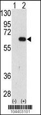
Western blot analysis of AMFR (arrow) using AMFR Antibody (N-term) . 293 cell lysates (2 ug/lane) either nontransfected (Lane 1) or transiently transfected with the AMFR gene (Lane 2) (Origene Technologies).Application
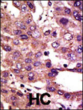
Formalin-fixed and paraffin-embedded human cancer tissue reacted with the primary antibody, which was peroxidase-conjugated to the secondary antibody, followed by DAB staining. This data demonstrates the use of this antibody for immunohistochemistry; clinical relevance has not been evaluated. BC = breast carcinoma; HC = hepatocarcinoma.Application
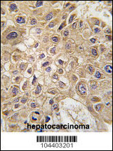
Formalin-fixed and paraffin-embedded human hepatocarcinoma tissue reacted with AMFR Antibody (N-term) , which was peroxidase-conjugated to the secondary antibody, followed by DAB staining. This data demonstrates the use of this antibody for immunohistochemistry; clinical relevance has not been evaluated.Application
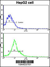
Flow cytometric analysis of HepG2 cells using AMFR Antibody (N-term)(bottom histogram) compared to a negative control cell (top histogram). FITC-conjugated goat-anti-rabbit secondary antibodies were used for the analysis.| Product Name | AMFR Antibody (N-term) |
|---|---|
| Antibody Type | Primary Antibodies |
| Antigen Alias | AMFR; RNF45; E3 ubiquitin-protein ligase AMFR; Autocrine motility factor receptor, isoform 2; RING finger protein 45; gp78 |
| Clonality | Polyclonal |
|---|---|
| Isotype | Ig |
| Host Species | Rabbit |
| Tested Applications | WBIHCFC |
| WB:1:100~500 IHC |
|
| Species Reactivity | Human |
| Concentration | 1mg/ml |
| Gene Synonyms | RNF45 |
|---|---|
| Alternative Names | AMFR RNF45 E3 ubiquitin-protein ligase AMFR Autocrine motility factor receptor isoform 2 RING finger protein 45 gp78 |
| Molecular Weight(MW) | 72996 Da |
| Function | E3 ubiquitin-protein ligase that mediates the polyubiquitination of a number of proteins such as CD3D, CYP3A4, CFTR and APOB for proteasomal degradation. Component of a VCP/p97- AMFR/gp78 complex that participates in the final step of endoplasmic reticulum-associated degradation (ERAD). The VCP/p97- AMFR/gp78 complex is involved in the sterol-accelerated ERAD degradation of HMGCR through binding to the HMGCR-INSIG complex at the ER membrane and initiating ubiquitination of HMGCR. The ubiquitinated HMGCR is then released from the ER by the complex into the cytosol for subsequent destruction. Also acts as a scaffold protein to assemble a complex that couples ubiquitination, retranslocation and deglycosylation. Mediates tumor invasion and metastasis as a receptor for the GPI/autocrine motility factor |
| Tissue Specificity | This AMFR antibody is generated from rabbits immunized with a KLH conjugated synthetic peptide between 4~33 amino acids from the N-terminal region of human AMFR. |
| Cellular Localization | Endoplasmic reticulum membrane; Multi-pass membrane protein |
Application
Western blot analysis of AMFR (arrow) using AMFR Antibody (N-term) . 293 cell lysates (2 ug/lane) either nontransfected (Lane 1) or transiently transfected with the AMFR gene (Lane 2) (Origene Technologies).
Application
Formalin-fixed and paraffin-embedded human cancer tissue reacted with the primary antibody, which was peroxidase-conjugated to the secondary antibody, followed by DAB staining. This data demonstrates the use of this antibody for immunohistochemistry; clinical relevance has not been evaluated. BC = breast carcinoma; HC = hepatocarcinoma.
Application
Formalin-fixed and paraffin-embedded human hepatocarcinoma tissue reacted with AMFR Antibody (N-term) , which was peroxidase-conjugated to the secondary antibody, followed by DAB staining. This data demonstrates the use of this antibody for immunohistochemistry; clinical relevance has not been evaluated.
Application
Flow cytometric analysis of HepG2 cells using AMFR Antibody (N-term)(bottom histogram) compared to a negative control cell (top histogram). FITC-conjugated goat-anti-rabbit secondary antibodies were used for the analysis.| Application Notes | WB:1:100~500 IHC |
|---|
| Form | Liquid |
|---|---|
| Storage Instructions | For short-term storage, store at 4° C. For long-term storage, aliquot and store at -20ºC or below. Avoid multiple freeze-thaw cycles. |
| Storage Buffer | Purified polyclonal antibody supplied in PBS with 0.09% (W/V) sodium azide. This antibody is purified through a protein G column, eluted with high and low pH buffers and neutralized immediately, followed by dialysis against PBS. |


