Application
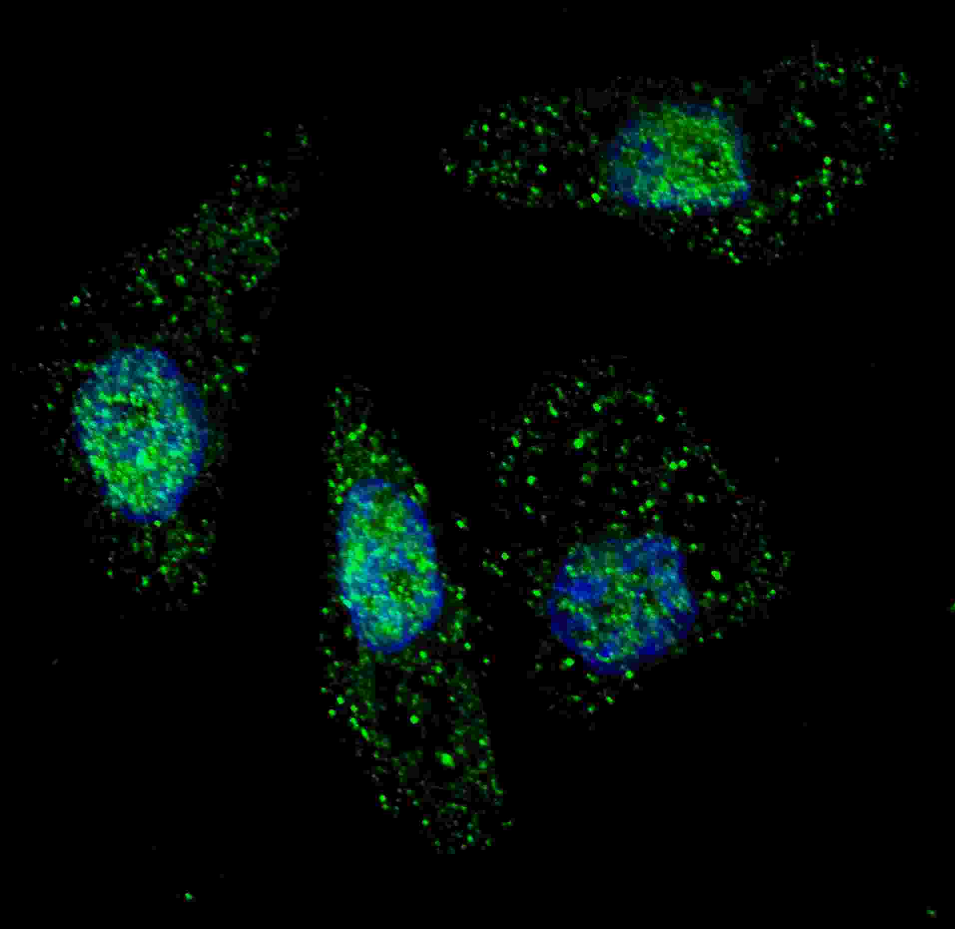
Fluorescent image of U251 cells stained with ATG7 antibody. U251 cells were treated with Chloroquine (50 μM,16h), then fixed with 4% PFA (20 min), permeabilized with Triton X-100 (0.2%, 30 min). Cells were then incubated with AP1813c ATG7 primary antibody (1:200, 2 h at room temperature). For secondary antibody, Alexa Fluor® 488 conjugated donkey anti-rabbit antibody (green) was used (1:1000, 1h). Nuclei were counterstained with Hoechst 33342 (blue) (10 μg/ml, 5 min). ATG7 immunoreactivity is localized to autophagic vacuoles in the cytoplasm of U251 cells, supported by Human Protein Atlas Data (http://www.proteinatlas.org/ENSG00000197548).Application
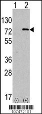
Western blot analysis of anti-hAPG7L-R509 Pab in 293 cell line lysates transiently transfected with the ATG7 gene (2ug/lane). hAPG7L-R509(arrow) was detected using the purified Pab (1:60 dilution).Application
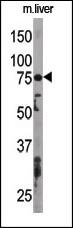
The anti-APG7L Pab is used in Western blot to detect APG7L in mouse liver tissue lysate. APG7L(arrow) was detected using the purified Pab.Application
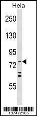
APG7L Antibody (R509) western blot analysis in Hela cell line lysates (35ug/lane).This demonstrates the APG7L antibody detected the APG7L protein (arrow).Application
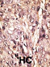
Formalin-fixed and paraffin-embedded human cancer tissue reacted with the primary antibody, which was peroxidase-conjugated to the secondary antibody, followed by AEC staining. This data demonstrates the use of this antibody for immunohistochemistry; clinical relevance has not been evaluated. BC = breast carcinoma; HC = hepatocarcinoma.| Product Name | ATG7 Antibody |
|---|---|
| Antibody Type | Primary Antibodies |
| Clonality | Polyclonal |
|---|---|
| Isotype | Ig |
| Host Species | Rabbit |
| Tested Applications | IFWBIHC |
| IF:1:200 WB |
|
| Species Reactivity | Human |
| Concentration | 1mg/ml |
| Gene Synonyms | APG7L |
|---|---|
| Alternative Names | ATG7 APG7L Ubiquitin-like modifier-activating enzyme ATG7 ATG12-activating enzyme E1 ATG7 Autophagy-related protein 7 Ubiquitin-activating enzyme E1-like protein |
| Molecular Weight(MW) | 77960 Da |
| Function | Functions as an E1 enzyme essential for multisubstrates such as ATG8-like proteins and ATG12. Forms intermediate conjugates with ATG8-like proteins (GABARAP, GABARAPL1, GABARAPL2 or MAP1LC3A). PE-conjugation to ATG8-like proteins is essential for autophagy. Also acts as an E1 enzyme for ATG12 conjugation to ATG5 and ATG3 (By similarity) |
| Tissue Specificity | Widely expressed, especially in kidney, liver, lymph nodes and bone marrow |
| Cellular Localization | Cytoplasm (Probable). |
| Entrez Gene | 10533 |
|---|
Application
Fluorescent image of U251 cells stained with ATG7 antibody. U251 cells were treated with Chloroquine (50 μM,16h), then fixed with 4% PFA (20 min), permeabilized with Triton X-100 (0.2%, 30 min). Cells were then incubated with AP1813c ATG7 primary antibody (1:200, 2 h at room temperature). For secondary antibody, Alexa Fluor® 488 conjugated donkey anti-rabbit antibody (green) was used (1:1000, 1h). Nuclei were counterstained with Hoechst 33342 (blue) (10 μg/ml, 5 min). ATG7 immunoreactivity is localized to autophagic vacuoles in the cytoplasm of U251 cells, supported by Human Protein Atlas Data (http://www.proteinatlas.org/ENSG00000197548).
Application
Western blot analysis of anti-hAPG7L-R509 Pab in 293 cell line lysates transiently transfected with the ATG7 gene (2ug/lane). hAPG7L-R509(arrow) was detected using the purified Pab (1:60 dilution).
Application
The anti-APG7L Pab is used in Western blot to detect APG7L in mouse liver tissue lysate. APG7L(arrow) was detected using the purified Pab.
Application
APG7L Antibody (R509) western blot analysis in Hela cell line lysates (35ug/lane).This demonstrates the APG7L antibody detected the APG7L protein (arrow).
Application
Formalin-fixed and paraffin-embedded human cancer tissue reacted with the primary antibody, which was peroxidase-conjugated to the secondary antibody, followed by AEC staining. This data demonstrates the use of this antibody for immunohistochemistry; clinical relevance has not been evaluated. BC = breast carcinoma; HC = hepatocarcinoma.| Application Notes | IF:1:200 WB |
|---|
| Form | Liquid |
|---|---|
| Storage Instructions | For short-term storage, store at 4° C. For long-term storage, aliquot and store at -20ºC or below. Avoid multiple freeze-thaw cycles. |
| Storage Buffer | Purified polyclonal antibody supplied in PBS with 0.09% (W/V) sodium azide. This antibody is prepared by Saturated Ammonium Sulfate (SAS) precipitation followed by dialysis against PBS. |


