Application
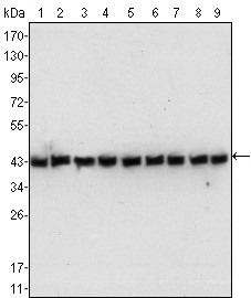
Figure 1: Western blot analysis using anti beta-Actin mAb against NIH/3T3 (1), Jurkat (2), Hela (3), CHO (4), PC12(5), HEK293 (6), COS (7), A549 (8) and MCF-7 (9) cell lysate.Application
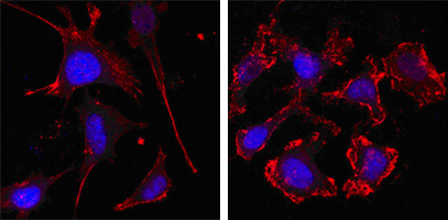
Figure 3: Confocal immunofluorescence analysis of SKBR-3 (left) and A549 (right) cells using anti-beta Actin mAb (red, the secondary Ab is Cy3-Goat anti mouse IgG). Blue: DRAQ5 fluorescent DNA dye.Application
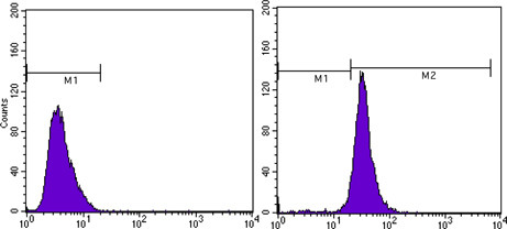
Figure 4: Flow cytometric analysis of MCF-7 cells using anti-beta Actin mAb (right) and negative control (left).| Product Name | beta-Actin Antibody |
|---|---|
| Antibody Type | Primary Antibodies |
| Antigen Alias | PS1TP5BP1; ACTB |
| Clonality | Monoclonal |
|---|---|
| Isotype | IgG2b |
| Clone Number | 8H10D10 |
| Host Species | Mouse |
| Tested Applications | WBIFFC |
| WB:1:500 - 2000. IF |
|
| Species Reactivity | Human |
| Concentration | 1mg/ml |
| Alternative Names | PS1TP5BP1 ACTB |
|---|---|
| Molecular Weight(MW) | 41737 Da |
| Function | Actins are highly conserved proteins that are involved in various types of cell motility and are ubiquitously expressed in all eukaryotic cells |
| Tissue Specificity | Synthetic peptide corresponding to amino-terminal residues of human beta-Actin, conjugated to KLH. |
| Cellular Localization | Cytoplasm, cytoskeleton. Note=Localized in cytoplasmic mRNP granules containing untranslated mRNAs |
| Entrez Gene | 60 |
|---|---|
| Nucleotide Accession | NP_001092.1 |
Application
Figure 1: Western blot analysis using anti beta-Actin mAb against NIH/3T3 (1), Jurkat (2), Hela (3), CHO (4), PC12(5), HEK293 (6), COS (7), A549 (8) and MCF-7 (9) cell lysate.
Application
Figure 3: Confocal immunofluorescence analysis of SKBR-3 (left) and A549 (right) cells using anti-beta Actin mAb (red, the secondary Ab is Cy3-Goat anti mouse IgG). Blue: DRAQ5 fluorescent DNA dye.
Application
Figure 4: Flow cytometric analysis of MCF-7 cells using anti-beta Actin mAb (right) and negative control (left).| Application Notes | WB:1:500 - 2000. IF |
|---|
| Form | Liquid |
|---|---|
| Storage Instructions | For short-term storage, store at 4° C. For long-term storage, aliquot and store at -20ºC or below. Avoid multiple freeze-thaw cycles. |
| Storage Buffer | Synthetic peptide corresponding to amino-terminal residues of human beta-Actin, conjugated to KLH. |


