Application
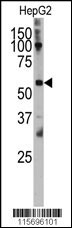
Western blot analysis of anti-CAMK4 Antibody (Center H353) Pab in HepG2 cell line lysates (35ug/lane). CAMK4 (arrow) was detected using the purified Pab.Application
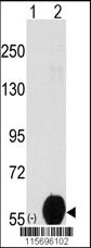
Western blot analysis of CAMK4 (Center H353) (arrow) using rabbit polyclonal CAMK4 (Center H353) Antibody . 293 cell lysates (2 ug/lane) either nontransfected (Lane 1) or transiently transfected with the CAMK4 (Center H353) gene (Lane 2)(Origene Technologies).Application
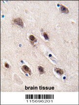
Formalin-fixed and paraffin-embedded human brain tissue reacted with CAMK4 (Center H353), which was peroxidase-conjugated to the secondary antibody, followed by DAB staining. This data demonstrates the use of this antibody for immunohistochemistry; clinical relevance has not been evaluated.| Product Name | CAMK4 Antibody (Center H353) |
|---|---|
| Antibody Type | Primary Antibodies |
| Antigen Alias | CAMK4; CAMK; CAMK-GR; CAMKIV; Calcium/calmodulin-dependent protein kinase type IV; CaM kinase-GR |
| Clonality | Polyclonal |
|---|---|
| Isotype | Ig |
| Host Species | Rabbit |
| Tested Applications | WBIHC |
| WB:1:50~100 IHC |
|
| Species Reactivity | Human |
| Concentration | 1mg/ml |
| Gene Synonyms | CAMK CAMK-GR CAMKIV |
|---|---|
| Alternative Names | CAMK4 CAMK CAMK-GR CAMKIV Calcium/calmodulin-dependent protein kinase type IV CaM kinase-GR |
| Molecular Weight(MW) | 51926 Da |
| Function | Calcium/calmodulin-dependent protein kinase that operates in the calcium-triggered CaMKK-CaMK4 signaling cascade and regulates, mainly by phosphorylation, the activity of several transcription activators, such as CREB1, MEF2D, JUN and RORA, which play pivotal roles in immune response, inflammation, and memory consolidation. In the thymus, regulates the CD4(+)/CD8(+) double positive thymocytes selection threshold during T-cell ontogeny. In CD4 memory T-cells, is required to link T-cell antigen receptor (TCR) signaling to the production of IL2, IFNG and IL4 (through the regulation of CREB and MEF2). Regulates the differentiation and survival phases of osteoclasts and dendritic cells (DCs). Mediates DCs survival by linking TLR4 and the regulation of temporal expression of BCL2. Phosphorylates the transcription activator CREB1 on 'Ser-133' in hippocampal neuron nuclei and contribute to memory consolidation and long term potentiation (LTP) in the hippocampus. Can activate the MAP kinases MAPK1/ERK2, MAPK8/JNK1 and MAPK14/p38 and stimulate transcription through the phosphorylation of ELK1 and ATF2. Can also phosphorylate in vitro CREBBP, PRM2, MEF2A and STMN1/OP18 |
| Tissue Specificity | Expressed in brain, thymus, CD4 T-cells, testis and epithelial ovarian cancer tissue |
| Cellular Localization | Cytoplasm. Nucleus. Note=Localized in hippocampal neuron nuclei. In spermatids, associated with chromatin and nuclear matrix (By similarity) |
| Entrez Gene | 814 |
|---|
Application
Western blot analysis of anti-CAMK4 Antibody (Center H353) Pab in HepG2 cell line lysates (35ug/lane). CAMK4 (arrow) was detected using the purified Pab.
Application
Western blot analysis of CAMK4 (Center H353) (arrow) using rabbit polyclonal CAMK4 (Center H353) Antibody . 293 cell lysates (2 ug/lane) either nontransfected (Lane 1) or transiently transfected with the CAMK4 (Center H353) gene (Lane 2)(Origene Technologies).
Application
Formalin-fixed and paraffin-embedded human brain tissue reacted with CAMK4 (Center H353), which was peroxidase-conjugated to the secondary antibody, followed by DAB staining. This data demonstrates the use of this antibody for immunohistochemistry; clinical relevance has not been evaluated.| Application Notes | WB:1:50~100 IHC |
|---|
| Form | Liquid |
|---|---|
| Storage Instructions | For short-term storage, store at 4° C. For long-term storage, aliquot and store at -20ºC or below. Avoid multiple freeze-thaw cycles. |
| Storage Buffer | Purified polyclonal antibody supplied in PBS with 0.09% (W/V) sodium azide. This antibody is prepared by Saturated Ammonium Sulfate (SAS) precipitation followed by dialysis against PBS. |


