Application
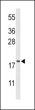
Western blot analysis of CFL1 Antibody (Center) in HL-60 cell line lysates (35ug/lane). CFL1 (arrow) was detected using the purified Pab;Application
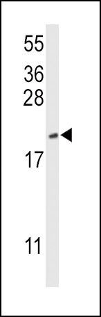
Western blot analysis of CFL1 Antibody (Center) in mouse NIH-3T3 cell line lysates (35ug/lane). CFL1 (arrow) was detected using the purified Pab.Application
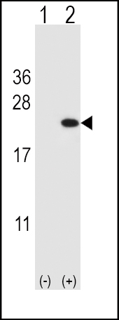
Western blot analysis of CFL1 (arrow) using rabbit polyclonal CFL1 Antibody (Center) . 293 cell lysates (2 ug/lane) either nontransfected (Lane 1) or transiently transfected (Lane 2) with the CFL1 gene.Application
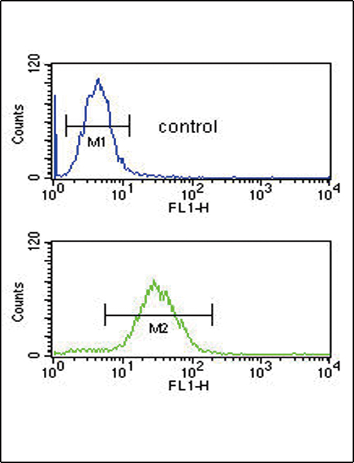
CFL1 Antibody (Center) flow cytometric analysis of HL-60 cells (bottom histogram) compared to a negative control cell (top histogram).FITC-conjugated goat-anti-rabbit secondary antibodies were used for the analysis.| Product Name | CFL1 Antibody (Center) |
|---|---|
| Antibody Type | Primary Antibodies |
| Antigen Alias | CFL1; CFL; Cofilin-1; 18 kDa phosphoprotein; Cofilin, non-muscle isoform |
| Clonality | Polyclonal |
|---|---|
| Isotype | Ig |
| Host Species | Rabbit |
| Tested Applications | WBFC |
| WB:1:100~500 FC |
|
| Species Reactivity | Human |
| Concentration | 1mg/ml |
| Gene Synonyms | CFL |
|---|---|
| Alternative Names | CFL1 CFL Cofilin-1 18 kDa phosphoprotein Cofilin non-muscle isoform |
| Molecular Weight(MW) | 18502 Da |
| Function | Binds to F-actin and exhibits pH-sensitive F-actin depolymerizing activity. Regulates actin cytoskeleton dynamics Important for normal progress through mitosis and normal cytokinesis. Plays a role in the regulation of cell morphology and cytoskeletal organization |
| Tissue Specificity | Widely distributed in various tissues. |
| Cellular Localization | Nucleus matrix. Cytoplasm, cytoskeleton. Cell projection, ruffle membrane; Peripheral membrane protein; Cytoplasmic side. Cell projection, lamellipodium membrane; Peripheral membrane protein; Cytoplasmic side. Note=Colocalizes with the actin cytoskeleton in membrane ruffles and lamellipodia Detected at the cleavage furrow and contractile ring during cytokinesis. Almost completely in nucleus in cells exposed to heat shock or 10% dimethyl sulfoxide |
| Entrez Gene | 1072 |
|---|
Application
Western blot analysis of CFL1 Antibody (Center) in HL-60 cell line lysates (35ug/lane). CFL1 (arrow) was detected using the purified Pab;
Application
Western blot analysis of CFL1 Antibody (Center) in mouse NIH-3T3 cell line lysates (35ug/lane). CFL1 (arrow) was detected using the purified Pab.
Application
Western blot analysis of CFL1 (arrow) using rabbit polyclonal CFL1 Antibody (Center) . 293 cell lysates (2 ug/lane) either nontransfected (Lane 1) or transiently transfected (Lane 2) with the CFL1 gene.
Application
CFL1 Antibody (Center) flow cytometric analysis of HL-60 cells (bottom histogram) compared to a negative control cell (top histogram).FITC-conjugated goat-anti-rabbit secondary antibodies were used for the analysis.| Application Notes | WB:1:100~500 FC |
|---|
| Form | Liquid |
|---|---|
| Storage Instructions | For short-term storage, store at 4° C. For long-term storage, aliquot and store at -20ºC or below. Avoid multiple freeze-thaw cycles. |
| Storage Buffer | Purified polyclonal antibody supplied in PBS with 0.09% (W/V) sodium azide. This antibody is purified through a protein A column, followed by peptide affinity purification. |


