Application
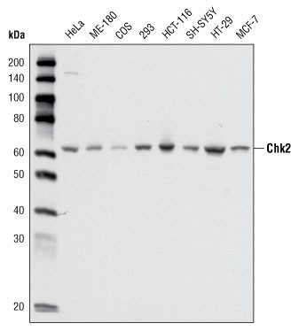
Figure 1: Western blot analysis using anti-CHK2 mAb against cell lysate from various cell types.Application
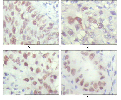
Figure 2: Immunohistochemical analysis of paraffin-embedded human lung carcinoma (A), liver carcinoma (B), breast carcinoma (C) and kiney carcinoma (D), showing nuclear localization with DAB staining using anti-CHK2 mAb.Application
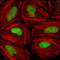
Figure 3: Confocal immunofluorescence analysis of Hela cells using anti-CHK2 mAb (green), showing nuclear localization. Red: Actin filaments have been labeled with DY-554 phalloidin.| Product Name | CHK2 Antibody |
|---|---|
| Antibody Type | Primary Antibodies |
| Antigen Alias | CDS1; LFS2; CHEK2 |
| Clonality | Monoclonal |
|---|---|
| Isotype | IgG2b |
| Clone Number | 1C12B8 |
| Host Species | Mouse |
| Tested Applications | WBIHCIF |
| WB:1:500 - 2000. IHC |
|
| Species Reactivity | Human |
| Concentration | 1mg/ml |
| Gene Synonyms | CDS1 CHK2 RAD53 |
|---|---|
| Alternative Names | CDS1 LFS2 CHEK2 |
| Molecular Weight(MW) | 60915 Da |
| Function | Serine/threonine-protein kinase which is required for checkpoint-mediated cell cycle arrest, activation of DNA repair and apoptosis in response to the presence of DNA double-strand breaks. May also negatively regulate cell cycle progression during unperturbed cell cycles. Following activation, phosphorylates numerous effectors preferentially at the consensus sequence [L-X- R-X-X-S/T]. Regulates cell cycle checkpoint arrest through phosphorylation of CDC25A, CDC25B and CDC25C, inhibiting their activity. Inhibition of CDC25 phosphatase activity leads to increased inhibitory tyrosine phosphorylation of CDK-cyclin complexes and blocks cell cycle progression. May also phosphorylate NEK6 which is involved in G2/M cell cycle arrest Regulates DNA repair through phosphorylation of BRCA2, enhancing the association of RAD51 with chromatin which promotes DNA repair by homologous recombination. Also stimulates the transcription of genes involved in DNA repair (including BRCA2) through the phosphorylation and activation of the transcription factor FOXM1 Regulates apoptosis through the phosphorylation of p53/TP53, MDM4 and PML. Phosphorylation of p53/TP53 at 'Ser-20' by CHEK2 may alleviate inhibition by MDM2, leading to accumulation of active p53/TP53. Phosphorylation of MDM4 may also reduce degradation of p53/TP53. Also controls the transcription of pro-apoptotic genes through phosphorylation of the transcription factor E2F1. Tumor suppressor, it may also have a DNA damage-independent function in mitotic spindle assembly by phosphorylating BRCA1. Its absence may be a cause of the chromosomal instability observed in some cancer cells |
| Tissue Specificity | High expression is found in testis, spleen, colon and peripheral blood leukocytes. Low expression is found in other tissues |
| Cellular Localization | Isoform 2: Nucleus. Note=Isoform 10 is present throughout the cell Isoform 7: Nucleus. Isoform 12: Nucleus. |
| Entrez Gene | 11200 |
|---|---|
| Nucleotide Accession | NP_665861.1 |
Application
Figure 1: Western blot analysis using anti-CHK2 mAb against cell lysate from various cell types.
Application
Figure 2: Immunohistochemical analysis of paraffin-embedded human lung carcinoma (A), liver carcinoma (B), breast carcinoma (C) and kiney carcinoma (D), showing nuclear localization with DAB staining using anti-CHK2 mAb.
Application
Figure 3: Confocal immunofluorescence analysis of Hela cells using anti-CHK2 mAb (green), showing nuclear localization. Red: Actin filaments have been labeled with DY-554 phalloidin.| Application Notes | WB:1:500 - 2000. IHC |
|---|
| Form | Liquid |
|---|---|
| Storage Instructions | For short-term storage, store at 4° C. For long-term storage, aliquot and store at -20ºC or below. Avoid multiple freeze-thaw cycles. |
| Storage Buffer | Purified recombinant fragment of human CHK2 (aa481-531) expressed in E. Coli. |


