Application
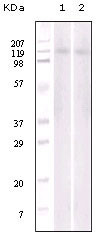
Figure 1: Western blot analysis of Jurkat (1) and NIH/3T3 (2) cell lysate using anti-EphB6 antibody.Application
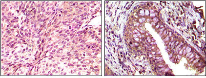
Figure 2: Immunohistochemical analysis of paraffin-embedded human bladder carcinoma(left) and return carcinoma(right) tissue, showing cytoplasmic localization using anti-EphB6 antibody with DAB staining.| Product Name | EphB6 Antibody |
|---|---|
| Antibody Type | Primary Antibodies |
| Clonality | Monoclonal |
|---|---|
| Isotype | IgG1 |
| Clone Number | 8E7H12 |
| Host Species | Mouse |
| Tested Applications | WBIHC |
| WB:1:500 - 2000. IHC |
|
| Species Reactivity | Human |
| Concentration | 1mg/ml |
| Alternative Names | HEP MGC129910 MGC129911 |
|---|---|
| Molecular Weight(MW) | 110700 Da |
| Function | Kinase-defective receptor for members of the ephrin-B family. Binds to ephrin-B1 and ephrin-B2. Modulates cell adhesion and migration by exerting both positive and negative effects upon stimulation with ephrin-B2. Inhibits JNK activation, T-cell receptor-induced IL-2 secretion and CD25 expression upon stimulation with ephrin-B2 |
| Tissue Specificity | Expressed in brain. Expressed in non invasive breast carcinoma cell lines (at protein level). Strong expression in brain and pancreas, and weak expression in other tissues, such as heart, placenta, lung, liver, skeletal muscle and kidney Expressed in breast non invasive tumors but not in metastatic lesions. Isoform 3 is expressed in cell lines of glioblastomas, anaplastic astrocytomas, gliosarcomas and astrocytomas. Isoform 3 is not detected in normal tissues |
| Cellular Localization | Membrane; Single-pass type I membrane protein |
| Entrez Gene | 2051 |
|---|---|
| Nucleotide Accession | NP_004436.2 |
Application
Figure 1: Western blot analysis of Jurkat (1) and NIH/3T3 (2) cell lysate using anti-EphB6 antibody.
Application
Figure 2: Immunohistochemical analysis of paraffin-embedded human bladder carcinoma(left) and return carcinoma(right) tissue, showing cytoplasmic localization using anti-EphB6 antibody with DAB staining.| Application Notes | WB:1:500 - 2000. IHC |
|---|
| Form | Liquid |
|---|---|
| Storage Instructions | For short-term storage, store at 4° C. For long-term storage, aliquot and store at -20ºC or below. Avoid multiple freeze-thaw cycles. |
| Storage Buffer | Purified recombinant fragment of EphB6 expressed in E. Coli. |


