Application
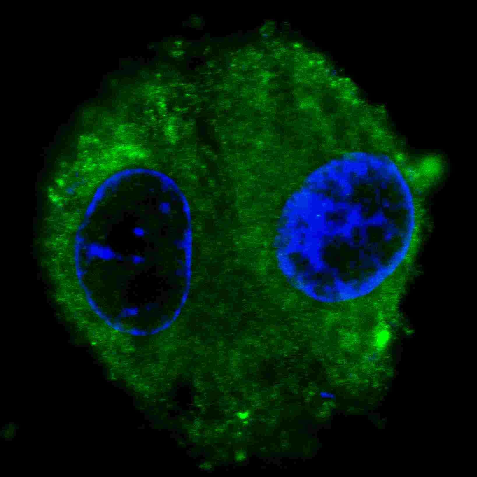
Fluorescent confocal image of HepG2 cells stained with FASN (Center) antibody. HepG2 cells were fixed with 4% PFA (20 min), permeabilized with Triton X-100 (0.2%, 30 min). Cells were then incubated with AM2067a FASN primary antibody (1:200, 2 h at room temperature). For secondary antibody, Alexa Fluor® 488 conjugated donkey anti-mouse antibody (green) was used (1:1000, 1h). Nuclei were counterstained with Hoechst 33342 (blue) (10 μg/ml, 5 min). Note the highly specific localization of the FASN immunosignal to the cytoplasm, supported by Human Protein Atlas Data (http://www.proteinatlas.org/ENSG00000169710).Application
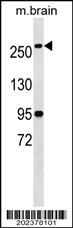
FASN Antibody (Center) (Cat. #AM2067a) western blot analysis in mouse brain tissue lysates (35ug/lane).This demonstrates the FASN antibody detected the FASN protein (arrow).| Product Name | FASN Antibody (Center) (Ascites) |
|---|---|
| Antibody Type | Primary Antibodies |
| Antigen Alias | FASN; FAS; Fatty acid synthase; [Acyl-carrier-protein] S-acetyltransferase; [Acyl-carrier-protein] S-malonyltransferase; 3-oxoacyl-[acyl-carrier-protein] synthase; 3-oxoacyl-[acyl-carrier-protein] reductase; 3-hydroxypalmitoyl-[acyl-carrier-protein] dehydr |
| Clonality | Monoclonal |
|---|---|
| Isotype | IgM |
| Clone Number | 497CT15.2.5 |
| Host Species | Mouse |
| Tested Applications | IFWB |
| IF:1:200 WB |
|
| Species Reactivity | Human |
| Concentration | 1mg/ml |
| Gene Synonyms | FAS |
|---|---|
| Alternative Names | FASN FAS Fatty acid synthase [Acyl-carrier-protein] S-acetyltransferase [Acyl-carrier-protein] S-malonyltransferase 3-oxoacyl-[acyl-carrier-protein] synthase 3-oxoacyl-[acyl-carrier-protein] reductase 3-hydroxypalmitoyl-[acyl-carrier-protein] dehydr |
| Molecular Weight(MW) | 273427 Da |
| Function | Fatty acid synthetase catalyzes the formation of long- chain fatty acids from acetyl-CoA, malonyl-CoA and NADPH. This multifunctional protein has 7 catalytic activities and an acyl carrier protein |
| Tissue Specificity | Ubiquitous. Prominent expression in brain, lung, and liver |
| Cellular Localization | Cytoplasm. Melanosome. Note=Identified by mass spectrometry in melanosome fractions from stage I to stage IV |
| Entrez Gene | 2194 |
|---|---|
| Nucleotide Accession | NP_004095.4 |
Application
Fluorescent confocal image of HepG2 cells stained with FASN (Center) antibody. HepG2 cells were fixed with 4% PFA (20 min), permeabilized with Triton X-100 (0.2%, 30 min). Cells were then incubated with AM2067a FASN primary antibody (1:200, 2 h at room temperature). For secondary antibody, Alexa Fluor® 488 conjugated donkey anti-mouse antibody (green) was used (1:1000, 1h). Nuclei were counterstained with Hoechst 33342 (blue) (10 μg/ml, 5 min). Note the highly specific localization of the FASN immunosignal to the cytoplasm, supported by Human Protein Atlas Data (http://www.proteinatlas.org/ENSG00000169710).
Application
FASN Antibody (Center) (Cat. #AM2067a) western blot analysis in mouse brain tissue lysates (35ug/lane).This demonstrates the FASN antibody detected the FASN protein (arrow).| Application Notes | IF:1:200 WB |
|---|
| Form | Liquid |
|---|---|
| Storage Instructions | For short-term storage, store at 4° C. For long-term storage, aliquot and store at -20ºC or below. Avoid multiple freeze-thaw cycles. |
| Storage Buffer | Mouse monoclonal antibody supplied in crude ascites with 0.09% (W/V) sodium azide. |


