Application
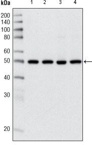
Figure 1: Western blot analysis using anti-HDAC3 mAb against Hela (1), NIH/3T3 (2), C6 (3) and COS (4) cell lysate.Application
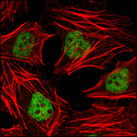
Figure 2: Confocal immunofluorescence analysis of Hela cells using anti-HDAC3 mAb (green). Red: Actin filaments have been labeled with DY-554 phalloidin.| Product Name | HDAC3 Antibody |
|---|---|
| Antibody Type | Primary Antibodies |
| Antigen Alias | HD3; RPD3; RPD3-2 |
| Clonality | Monoclonal |
|---|---|
| Isotype | IgG2a |
| Clone Number | 7G6C5 |
| Host Species | Mouse |
| Tested Applications | WBIF |
| WB:1:500 - 2000. IF |
|
| Species Reactivity | Human |
| Concentration | 1mg/ml |
| Alternative Names | HD3 RPD3 RPD3-2 |
|---|---|
| Molecular Weight(MW) | 48848 Da |
| Function | Responsible for the deacetylation of lysine residues on the N-terminal part of the core histones (H2A, H2B, H3 and H4), and some other non-histone substrates. Histone deacetylation gives a tag for epigenetic repression and plays an important role in transcriptional regulation, cell cycle progression and developmental events. Histone deacetylases act via the formation of large multiprotein complexes. Probably participates in the regulation of transcription through its binding to the zinc-finger transcription factor YY1; increases YY1 repression activity Required to repress transcription of the POU1F1 transcription factor. Acts as a molecular chaperone for shuttling phosphorylated NR2C1 to PML bodies for sumoylation (By similarity) |
| Tissue Specificity | Widely expressed. |
| Cellular Localization | Nucleus. |
| Entrez Gene | 8841 |
|---|---|
| Nucleotide Accession | NP_003874.2 |
Application
Figure 1: Western blot analysis using anti-HDAC3 mAb against Hela (1), NIH/3T3 (2), C6 (3) and COS (4) cell lysate.
Application
Figure 2: Confocal immunofluorescence analysis of Hela cells using anti-HDAC3 mAb (green). Red: Actin filaments have been labeled with DY-554 phalloidin.| Application Notes | WB:1:500 - 2000. IF |
|---|
| Form | Liquid |
|---|---|
| Storage Instructions | For short-term storage, store at 4° C. For long-term storage, aliquot and store at -20ºC or below. Avoid multiple freeze-thaw cycles. |
| Storage Buffer | Purified recombinant fragment of human HDAC3 (aa224-428) expressed in E. Coli. |


