Application
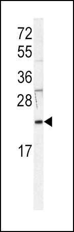
Western blot analysis of anti-HRAS Antibody (Center) in Jurkat cell line lysates (35ug/lane). HRAS (arrow) was detected using the purified Pab.Application
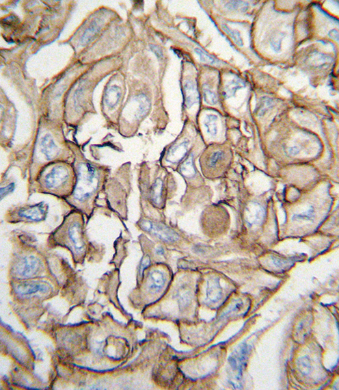
Formalin-fixed and paraffin-embedded human lung carcinoma tissue reacted with HRAS antibody (Center), which was peroxidase-conjugated to the secondary antibody, followed by DAB staining. This data demonstrates the use of this antibody for immunohistochemistry; clinical relevance has not been evaluated.Application
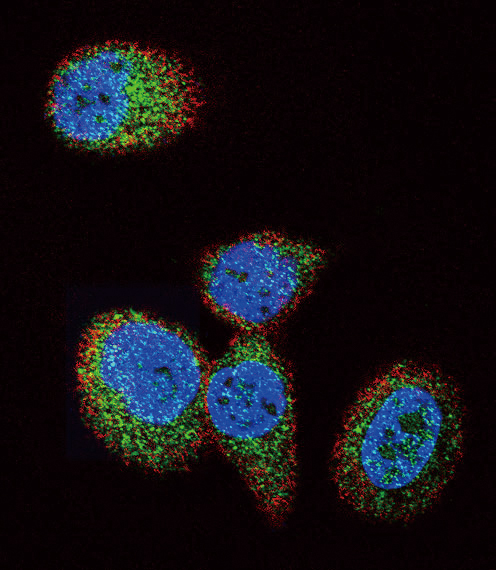
Confocal immunofluorescent analysis of HRAS Antibody (Center)(Cat#AP7764c) with MCF-7 cell followed by Alexa Fluor 488-conjugated goat anti-rabbit lgG (green).Actin filaments have been labeled with Alexa Fluor 555 phalloidin (red).DAPI was used to stain the cell nuclear (blue).| Product Name | HRAS Antibody (Center) |
|---|---|
| Antibody Type | Primary Antibodies |
| Antigen Alias | HRAS; HRAS1; GTPase HRas; H-Ras-1; Ha-Ras; Transforming protein p21; c-H-ras; p21ras; GTPase HRas, N-terminally processed |
| Clonality | Polyclonal |
|---|---|
| Isotype | Ig |
| Host Species | Rabbit |
| Tested Applications | WBIHCIF |
| WB:1:100~500 IHC |
|
| Species Reactivity | Human |
| Concentration | 1mg/ml |
| Gene Synonyms | HRAS1 |
|---|---|
| Alternative Names | HRAS HRAS1 GTPase HRas H-Ras-1 Ha-Ras Transforming protein p21 c-H-ras p21ras GTPase HRas N-terminally processed |
| Molecular Weight(MW) | 21298 Da |
| Function | Ras proteins bind GDP/GTP and possess intrinsic GTPase activity |
| Tissue Specificity | Widely expressed. |
| Cellular Localization | Cell membrane. Cell membrane; Lipid-anchor; Cytoplasmic side. Golgi apparatus. Golgi apparatus membrane; Lipid-anchor. Note=The active GTP-bound form is localized most strongly to membranes than the inactive GDP-bound form (By similarity). Shuttles between the plasma membrane and the Golgi apparatus |
| Entrez Gene | 3265 |
|---|
Application
Western blot analysis of anti-HRAS Antibody (Center) in Jurkat cell line lysates (35ug/lane). HRAS (arrow) was detected using the purified Pab.
Application
Formalin-fixed and paraffin-embedded human lung carcinoma tissue reacted with HRAS antibody (Center), which was peroxidase-conjugated to the secondary antibody, followed by DAB staining. This data demonstrates the use of this antibody for immunohistochemistry; clinical relevance has not been evaluated.
Application
Confocal immunofluorescent analysis of HRAS Antibody (Center)(Cat#AP7764c) with MCF-7 cell followed by Alexa Fluor 488-conjugated goat anti-rabbit lgG (green).Actin filaments have been labeled with Alexa Fluor 555 phalloidin (red).DAPI was used to stain the cell nuclear (blue).| Application Notes | WB:1:100~500 IHC |
|---|
| Form | Liquid |
|---|---|
| Storage Instructions | For short-term storage, store at 4° C. For long-term storage, aliquot and store at -20ºC or below. Avoid multiple freeze-thaw cycles. |
| Storage Buffer | Purified polyclonal antibody supplied in PBS with 0.09% (W/V) sodium azide. This antibody is prepared by Saturated Ammonium Sulfate (SAS) precipitation followed by dialysis against PBS. |


