Application
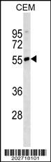
IGHD Antibody (N-term)(Ascites)(Cat. #AM2141a) western blot analysis in CEM cell line lysates (35ug/lane).This demonstrates the IGHD antibody detected the IGHD protein (arrow).| Product Name | IGHD Antibody (N-term) (Ascites) |
|---|---|
| Antibody Type | Primary Antibodies |
| Antigen Alias | IGHD; Ig delta chain C region |
| Clonality | Monoclonal |
|---|---|
| Isotype | IgM |
| Clone Number | 607CT8.4.4 |
| Host Species | Mouse |
| Tested Applications | WB |
| WB:1:100~800 |
|
| Species Reactivity | Human |
| Concentration | 1mg/ml |
| Alternative Names | IGHD Ig delta chain C region |
|---|---|
| Molecular Weight(MW) | 42254 Da |
| Function | IgD is the major antigen receptor isotype on the surface of most peripheral B-cells, where it is coexpressed with IgM. The membrane-bound IgD (mIgD) induces the phosphorylation of CD79A and CD79B by the Src family of protein tyrosine kinases. Soluble IgD (sIgD) concentration in serum below those of IgG, IgA, and IgM but much higher than that of IgE. IgM and IgD molecules present on B cells have identical V regions and antigen-binding sites. After the antigen binds to the B-cell receptor, the secreted form sIgD is shut off. IgD is a potent inducer of TNF, IL1B, and IL1RN. IgD also induces release of IL6, IL10, and LIF from peripheral blood mononuclear cells. Monocytes seem to be the main producers of cytokines in vitro in the presence of IgD |
| Tissue Specificity | This IGHD Monoclonal antibody is generated from mouses immunized with a KLH conjugated synthetic peptide selected from human IGHD. |
| Cellular Localization | Isoform 1: Secreted. |
Application
IGHD Antibody (N-term)(Ascites)(Cat. #AM2141a) western blot analysis in CEM cell line lysates (35ug/lane).This demonstrates the IGHD antibody detected the IGHD protein (arrow).| Application Notes | WB:1:100~800 |
|---|
| Form | Liquid |
|---|---|
| Storage Instructions | For short-term storage, store at 4° C. For long-term storage, aliquot and store at -20ºC or below. Avoid multiple freeze-thaw cycles. |
| Storage Buffer | Mouse monoclonal antibody supplied in crude ascites with 0.09% (W/V) sodium azide. |


