Application
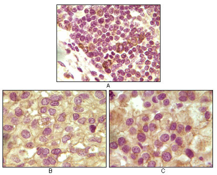
Figure 1: Immunohistochemical analysis of paraffin-embedded human lymphoid (A), ovary tumor (B) and testicle tumor (C) tissues using anti-INHA mAb with DAB staining.Application
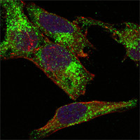
Figure 3: Confocal immunofluorescence analysis of Hela cells using anti-INHA mAb (green). Red: Actin filaments have been labeled with DY-554 phalloidin. Blue: DRAQ5 fluorescent DNA dye.| Product Name | INHA(Inhibin alpha) Antibody |
|---|---|
| Antibody Type | Primary Antibodies |
| Antigen Alias | INHA; inhibin, alpha |
| Clonality | Monoclonal |
|---|---|
| Isotype | IgG1 |
| Clone Number | 4A2 |
| Host Species | Mouse |
| Tested Applications | IHCIF |
| IHC: 1:200 - 1000. IF |
|
| Species Reactivity | Human |
| Concentration | 1mg/ml |
| Alternative Names | INHA inhibin alpha |
|---|---|
| Molecular Weight(MW) | 39670 Da |
| Function | Inhibins and activins inhibit and activate, respectively, the secretion of follitropin by the pituitary gland Inhibins/activins are involved in regulating a number of diverse functions such as hypothalamic and pituitary hormone secretion, gonadal hormone secretion, germ cell development and maturation, erythroid differentiation, insulin secretion, nerve cell survival, embryonic axial development or bone growth, depending on their subunit composition. Inhibins appear to oppose the functions of activins |
| Tissue Specificity | Originally found in ovary (granulosa cells) and testis (Sertoli cells), but widely distributed in many tissues including brain and placenta. In adrenal cortex expression is limited to the zona reticularis and the innermost zona fasciculata in the normal gland, extending centripetally into the zona fasciculata in hyperplasia. Also found in adrenocortical tumors Also expressed in prostate epithelium of benign prostatic hyperplasia, in regions of basal cell hyperplasia and in nonmalignant regions of high grade prostate cancer. Only circulating inhibin B is found in male, whereas circulating inhibins A and B are found in female |
| Cellular Localization | Secreted. |
| Entrez Gene | 3623 |
|---|---|
| Nucleotide Accession | NP_002182.1 |
Application
Figure 1: Immunohistochemical analysis of paraffin-embedded human lymphoid (A), ovary tumor (B) and testicle tumor (C) tissues using anti-INHA mAb with DAB staining.
Application
Figure 3: Confocal immunofluorescence analysis of Hela cells using anti-INHA mAb (green). Red: Actin filaments have been labeled with DY-554 phalloidin. Blue: DRAQ5 fluorescent DNA dye.| Application Notes | IHC: 1:200 - 1000. IF |
|---|
| Form | Liquid |
|---|---|
| Storage Instructions | For short-term storage, store at 4° C. For long-term storage, aliquot and store at -20ºC or below. Avoid multiple freeze-thaw cycles. |
| Storage Buffer | Purified recombinant fragment of human INHA expressed in E. Coli. |


