Application
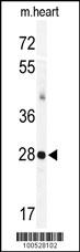
Western blot analysis of KChIP3 Antibody (N-term M1) (Cat.# AP1572a) in mouse heart tissue lysates (35ug/lane). KChIP3(arrow) was detected using the purified Pab.Application
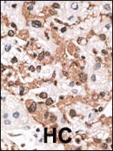
Formalin-fixed and paraffin-embedded human cancer tissue reacted with the primary antibody, which was peroxidase-conjugated to the secondary antibody, followed by AEC staining. This data demonstrates the use of this antibody for immunohistochemistry; clinical relevance has not been evaluated. BC = breast carcinoma; HC = hepatocarcinoma.Application
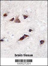
Formalin-fixed and paraffin-embedded human brain tissue reacted with KChIP3 Antibody (N-term M1) , which was peroxidase-conjugated to the secondary antibody, followed by DAB staining. This data demonstrates the use of this antibody for immunohistochemistry; clinical relevance has not been evaluated.Application
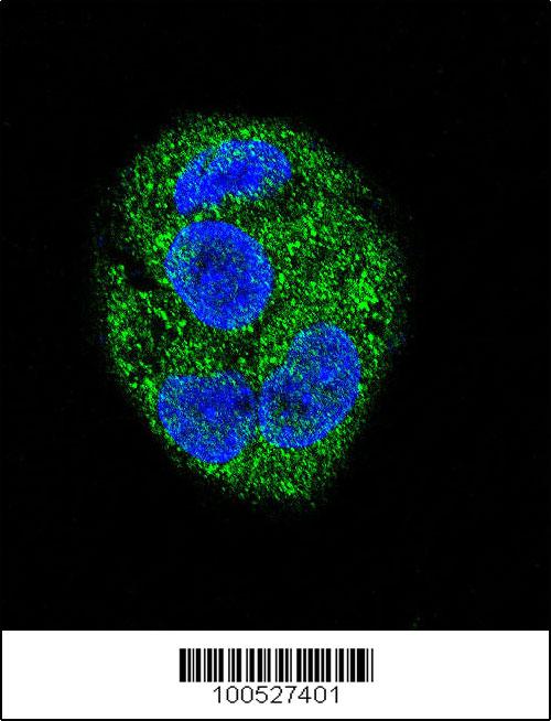
Confocal immunofluorescent analysis of KChIP3 Antibody (N-term M1) (Cat#AP1572a) with HepG2 cell followed by Alexa Fluor 488-conjugated goat anti-rabbit lgG (green). DAPI was used to stain the cell nuclear (blue).| Product Name | KChIP3 Antibody (N-term M1) |
|---|---|
| Antibody Type | Primary Antibodies |
| Antigen Alias | KCNIP3; CSEN; DREAM; KCHIP3; Calsenilin; A-type potassium channel modulatory protein 3; DRE-antagonist modulator; Kv channel-interacting protein 3 |
| Clonality | Polyclonal |
|---|---|
| Isotype | Ig |
| Host Species | Rabbit |
| Tested Applications | WBIHCIF |
| WB:1:100~500 IHC |
|
| Species Reactivity | Human |
| Concentration | 1mg/ml |
| Gene Synonyms | CSEN DREAM KCHIP3 |
|---|---|
| Alternative Names | KCNIP3 CSEN DREAM KCHIP3 Calsenilin A-type potassium channel modulatory protein 3 DRE-antagonist modulator Kv channel-interacting protein 3 |
| Molecular Weight(MW) | 29231 Da |
| Function | Calcium-dependent transcriptional repressor that binds to the DRE element of genes including PDYN and FOS. Affinity for DNA is reduced upon binding to calcium and enhanced by binding to magnesium. Seems to be involved in nociception (By similarity) May play a role in the regulation of PSEN2 proteolytic processing and apoptosis. Together with PSEN2 involved in modulation of beta-amyloid formation |
| Tissue Specificity | Highly expressed in brain. Widely expressed at lower levels. Expression levels are elevated in brain cortex regions affected by Alzheimer disease |
| Cellular Localization | Cytoplasm. Cell membrane; Lipid-anchor (By similarity). Endoplasmic reticulum. Golgi apparatus. Nucleus Note=Also membrane-bound, associated with the plasma membrane (By similarity). In the presence of PSEN2 associated with the endoplasmic reticulum and Golgi. The sumoylated form is present only in the nucleus |
| Entrez Gene | 30818 |
|---|
Application
Western blot analysis of KChIP3 Antibody (N-term M1) (Cat.# AP1572a) in mouse heart tissue lysates (35ug/lane). KChIP3(arrow) was detected using the purified Pab.
Application
Formalin-fixed and paraffin-embedded human cancer tissue reacted with the primary antibody, which was peroxidase-conjugated to the secondary antibody, followed by AEC staining. This data demonstrates the use of this antibody for immunohistochemistry; clinical relevance has not been evaluated. BC = breast carcinoma; HC = hepatocarcinoma.
Application
Formalin-fixed and paraffin-embedded human brain tissue reacted with KChIP3 Antibody (N-term M1) , which was peroxidase-conjugated to the secondary antibody, followed by DAB staining. This data demonstrates the use of this antibody for immunohistochemistry; clinical relevance has not been evaluated.
Application
Confocal immunofluorescent analysis of KChIP3 Antibody (N-term M1) (Cat#AP1572a) with HepG2 cell followed by Alexa Fluor 488-conjugated goat anti-rabbit lgG (green). DAPI was used to stain the cell nuclear (blue).| Application Notes | WB:1:100~500 IHC |
|---|
| Form | Liquid |
|---|---|
| Storage Instructions | For short-term storage, store at 4° C. For long-term storage, aliquot and store at -20ºC or below. Avoid multiple freeze-thaw cycles. |
| Storage Buffer | Purified polyclonal antibody supplied in PBS with 0.09% (W/V) sodium azide. This antibody is purified through a protein G column, eluted with high and low pH buffers and neutralized immediately, followed by dialysis against PBS. |


