Application
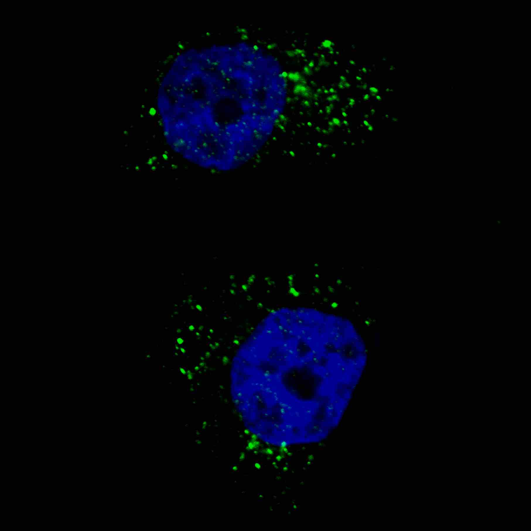
Fluorescent image of U251 cells stained with AM1800a LC3 (APG8) antibody.U251 cells were treated with Chloroquine (50 μM,16h), then fixed with 4% PFA (20 min), permeabilized with Triton X-100 (0.2%, 30 min). Cells were then incubated with AM1800a LC3 (APG8) primary antibody (1:200, 2 h at room temperature). For secondary antibody, Alexa Fluor® 488 conjugated donkey anti-mouse antibody (green) was used (1:1000, 1h). Nuclei were counterstained with Hoechst 33342 (blue) (10 μg/ml, 5 min). LC3 immunoreactivity is localized to autophagic vacuoles in the cytoplasm of U251 cells.Application
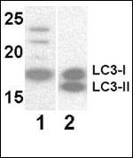
Western blot analysis of anti-LC3 Mab (Cat. #AM1800a) at 8 ug/ml. Lane 1: Y79 (soluble fraction of cell extract); Lane 2: 293 transfected with human LC3 (whole cell extract).Application
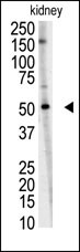
Western blot analysis of anti-LC3 Mab (Cat. #AM1800a) Hela cell lysates, which were treated with rapamycin or bafilomycin overnight. Data courtesy of Dr. David Rubinsztein, Cambridge Institute for Medical Research.Application
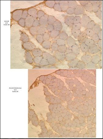
10X( lower panel) and 20X (upper panel) immunohistochemistry images from muscle tissue of a diseased mouse off Dox after 5 weeks on regular food. Several fibers that have autophagic vesicles throughout are visible. Primary antibody used is Cat# AM1800a. Data courtesy of Dr. Christy Caudill, Cincinnati Children's Hospital Medical Center.| Product Name | LC3 Antibody (APG8) |
|---|---|
| Antibody Type | Primary Antibodies |
| Antigen Alias | MAP1LC3A; Microtubule-associated proteins 1A/1B light chain 3A; Autophagy-related protein LC3 A; Autophagy-related ubiquitin-like modifier LC3 A; MAP1 light chain 3-like protein 1; MAP1A/MAP1B light chain 3 A; Microtubule-associated protein 1 light chain 3 |
| Clonality | Monoclonal |
|---|---|
| Isotype | IgG1 |
| Clone Number | 166AT1234 |
| Host Species | Mouse |
| Tested Applications | IFWBIHC |
| IF:1:200 WB |
|
| Species Reactivity | Human |
| Concentration | 1mg/ml |
| Alternative Names | MAP1LC3A Microtubule-associated proteins 1A/1B light chain 3A Autophagy-related protein LC3 A Autophagy-related ubiquitin-like modifier LC3 A MAP1 light chain 3-like protein 1 MAP1A/MAP1B light chain 3 A Microtubule-associated protein 1 light chain 3 |
|---|---|
| Molecular Weight(MW) | 14272 Da |
| Function | Involved in formation of autophagosomal vacuoles (autophagosomes) |
| Tissue Specificity | Most abundant in heart, brain, liver, skeletal muscle and testis but absent in thymus and peripheral blood leukocytes |
| Cellular Localization | Cytoplasm, cytoskeleton. Endomembrane system; Lipid-anchor. Cytoplasmic vesicle, autophagosome membrane; Lipid-anchor. Cytoplasmic vesicle, autophagosome. Note=LC3-II binds to the autophagic membranes |
| Entrez Gene | 84557 |
|---|
Application
Fluorescent image of U251 cells stained with AM1800a LC3 (APG8) antibody.U251 cells were treated with Chloroquine (50 μM,16h), then fixed with 4% PFA (20 min), permeabilized with Triton X-100 (0.2%, 30 min). Cells were then incubated with AM1800a LC3 (APG8) primary antibody (1:200, 2 h at room temperature). For secondary antibody, Alexa Fluor® 488 conjugated donkey anti-mouse antibody (green) was used (1:1000, 1h). Nuclei were counterstained with Hoechst 33342 (blue) (10 μg/ml, 5 min). LC3 immunoreactivity is localized to autophagic vacuoles in the cytoplasm of U251 cells.
Application
Western blot analysis of anti-LC3 Mab (Cat. #AM1800a) at 8 ug/ml. Lane 1: Y79 (soluble fraction of cell extract); Lane 2: 293 transfected with human LC3 (whole cell extract).
Application
Western blot analysis of anti-LC3 Mab (Cat. #AM1800a) Hela cell lysates, which were treated with rapamycin or bafilomycin overnight. Data courtesy of Dr. David Rubinsztein, Cambridge Institute for Medical Research.
Application
10X( lower panel) and 20X (upper panel) immunohistochemistry images from muscle tissue of a diseased mouse off Dox after 5 weeks on regular food. Several fibers that have autophagic vesicles throughout are visible. Primary antibody used is Cat# AM1800a. Data courtesy of Dr. Christy Caudill, Cincinnati Children's Hospital Medical Center.| Application Notes | IF:1:200 WB |
|---|
| Form | Liquid |
|---|---|
| Storage Instructions | For short-term storage, store at 4° C. For long-term storage, aliquot and store at -20ºC or below. Avoid multiple freeze-thaw cycles. |
| Storage Buffer | Purified monoclonal antibody supplied in PBS with 0.09% (W/V) sodium azide. This antibody is purified through a protein G column, eluted with high and low pH buffers and neutralized immediately, followed by dialysis against PBS. |


