Application
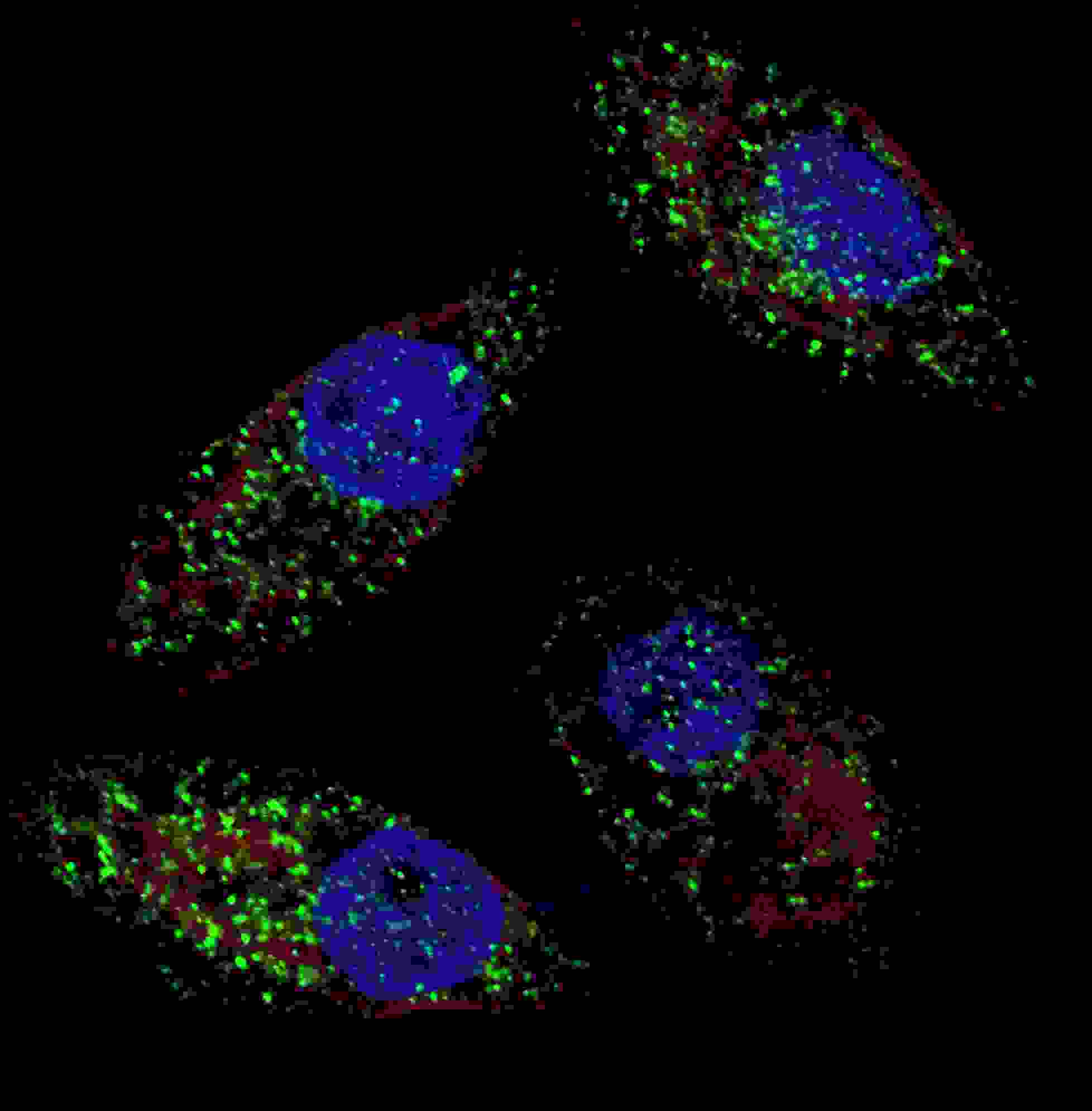
Fluorescent image of U251 cells stained with cleaved LC3B antibody. U251 cells were treated with Chloroquine (50 μM,16h), then fixed with 4% PFA (20 min), permeabilized with Triton X-100 (0.2%, 30 min). Cells were then incubated with AP1806a cleaved LC3B primary antibody (1:100, 2 h at room temperature). For secondary antibody, Alexa Fluor® 488 conjugated donkey anti-rabbit antibody (green) was used (1:1000, 1h). Nuclei were counterstained with Hoechst 33342 (blue) (10 μg/ml, 5 min). LC3 immunoreactivity is localized to autophagic vacuoles in the cytoplasm of U251 cells.Application
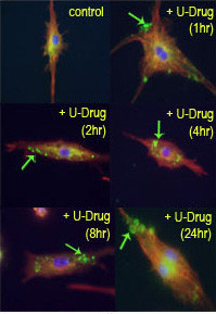
Time course study of mouse leukaemic monocyte macrophage cells treated with U18666A, a drug that causes cholesterol and lipid storage in cells, thereby blocking fusion between late endosomes and lysosomes. Cleaved-LC3 (APG8b) antibody (Cat AP1806a) detected punctuate staining indicative of autophagic vacuole or phagosome structures. Data courtesy of Dr. Barry Boland, Department of Pharmacology, Oxford University.| Product Name | Cleaved LC3B Antibody |
|---|---|
| Antibody Type | Primary Antibodies |
| Antigen Alias | MAP1LC3B; MAP1ALC3; Microtubule-associated proteins 1A/1B light chain 3B; Autophagy-related protein LC3 B; Autophagy-related ubiquitin-like modifier LC3 B; MAP1 light chain 3-like protein 2; MAP1A/MAP1B light chain 3 B; Microtubule-associated protein 1 lig |
| Clonality | Polyclonal |
|---|---|
| Isotype | Ig |
| Host Species | Rabbit |
| Tested Applications | IF |
| IF:1:10~50 |
|
| Species Reactivity | Human |
| Concentration | 1mg/ml |
| Gene Synonyms | MAP1ALC3 |
|---|---|
| Alternative Names | MAP1LC3B MAP1ALC3 Microtubule-associated proteins 1A/1B light chain 3B Autophagy-related protein LC3 B Autophagy-related ubiquitin-like modifier LC3 B MAP1 light chain 3-like protein 2 MAP1A/MAP1B light chain 3 B Microtubule-associated protein 1 lig |
| Molecular Weight(MW) | 14688 Da |
| Function | Involved in formation of autophagosomal vacuoles (autophagosomes) |
| Tissue Specificity | Most abundant in heart, brain, skeletal muscle and testis. Little expression observed in liver |
| Cellular Localization | Cytoplasm, cytoskeleton. Endomembrane system; Lipid-anchor. Cytoplasmic vesicle, autophagosome membrane; Lipid-anchor. Note=LC3-II binds to the autophagic membranes |
| Entrez Gene | 81631 |
|---|
Application
Fluorescent image of U251 cells stained with cleaved LC3B antibody. U251 cells were treated with Chloroquine (50 μM,16h), then fixed with 4% PFA (20 min), permeabilized with Triton X-100 (0.2%, 30 min). Cells were then incubated with AP1806a cleaved LC3B primary antibody (1:100, 2 h at room temperature). For secondary antibody, Alexa Fluor® 488 conjugated donkey anti-rabbit antibody (green) was used (1:1000, 1h). Nuclei were counterstained with Hoechst 33342 (blue) (10 μg/ml, 5 min). LC3 immunoreactivity is localized to autophagic vacuoles in the cytoplasm of U251 cells.
Application
Time course study of mouse leukaemic monocyte macrophage cells treated with U18666A, a drug that causes cholesterol and lipid storage in cells, thereby blocking fusion between late endosomes and lysosomes. Cleaved-LC3 (APG8b) antibody (Cat AP1806a) detected punctuate staining indicative of autophagic vacuole or phagosome structures. Data courtesy of Dr. Barry Boland, Department of Pharmacology, Oxford University.| Application Notes | IF:1:10~50 |
|---|
| Form | Liquid |
|---|---|
| Storage Instructions | For short-term storage, store at 4° C. For long-term storage, aliquot and store at -20ºC or below. Avoid multiple freeze-thaw cycles. |
| Storage Buffer | Purified polyclonal antibody supplied in PBS with 0.09% (W/V) sodium azide. This antibody is prepared by Saturated Ammonium Sulfate (SAS) precipitation followed by dialysis against PBS. |


