Application
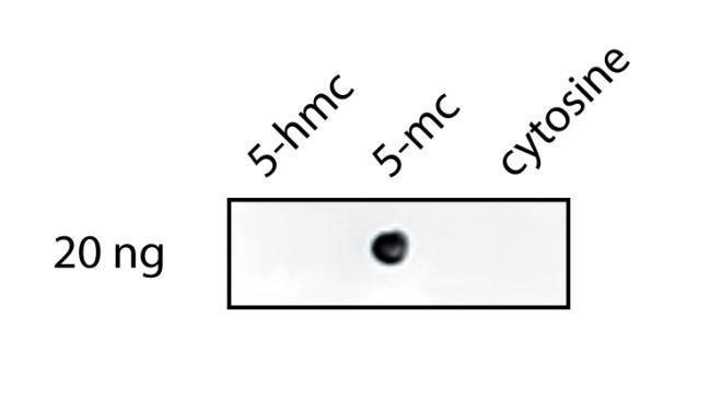
Dot ImageApplication
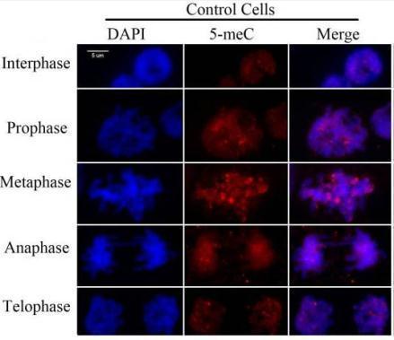
ICC/IF ImageImmunostaining for 5-meC in representative maize root tip cells in interphase or mitosis using 5-meC antibody . Pontes, O. et al. (2007) PLoS One. 2: e1157.Application
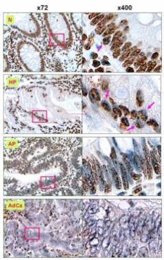
IHC Image5-MeC in DNA of various stages of colorectal lesions was detected by IHC staining using 5-MeC antibody . Sections of HPs, APs, AdCas and histologically “normal” mucosa around colorectal cancers were stained with mAb to 5-MeC and counterstained with hematoxylin. N (> 3 ~ 4+), “normal” mucosa ; HP (3+), the hyperplastic polyps; AP (2+), the adenomatous polyps; and AdCa (Application
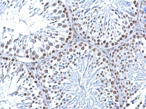
IHC-P Image5-Methylcytosine / 5-mC antibody [33D3] detects 5-Methylcytosine / 5-mC at nucleus on mouse testis by immunohistochemical analysis. Sample: Paraffin-embedded mouse testis. 5-Methylcytosine / 5-mC antibody [33D3] diluted at 1:200.| Product Name | 5-Methylcytosine / 5-mC antibody |
|---|---|
| Antibody Type | Primary Antibodies |
| Product description | Mouse monoclonal antibody [33D3] to 5-Methylcytosine / 5-mC |
| Clonality | Monoclonal |
|---|---|
| Isotype | IgG1 |
| Clone Number | 33D3 |
| Host Species | Mouse |
| Tested Applications | DotFACSICC/IFIHCIHC-FIHC-PIBMeDIP |
| FACS:1:200~1:500 ICC/IF:1:200~1:500 IHC:1:50~1:200 IHC-F:1:50~1:100 IHC-P:1:50~1:200 Notes:Optimal dilutions/concentrations should be determined by the researcher. |
|
| Species Reactivity | HumanMouseRatBovineAll |
| Concentration | 1mg/ml |
| Target | 5-Methylcytosine / 5-mC |
|---|---|
| Tissue Specificity | This antibody recognizes 5-Methylcytosine. Clone 33D3 is specific to the methylated base and has minimal reactivity to non-methylated cytidine or cytosine (Reynaudet al.1992). It has been reported for use in Methylated DNA immunoprecipitation (MeDIP) (Pon |
Application
Dot Image
Application
ICC/IF ImageImmunostaining for 5-meC in representative maize root tip cells in interphase or mitosis using 5-meC antibody . Pontes, O. et al. (2007) PLoS One. 2: e1157.
Application
IHC Image5-MeC in DNA of various stages of colorectal lesions was detected by IHC staining using 5-MeC antibody . Sections of HPs, APs, AdCas and histologically “normal” mucosa around colorectal cancers were stained with mAb to 5-MeC and counterstained with hematoxylin. N (> 3 ~ 4+), “normal” mucosa ; HP (3+), the hyperplastic polyps; AP (2+), the adenomatous polyps; and AdCa (
Application
IHC-P Image5-Methylcytosine / 5-mC antibody [33D3] detects 5-Methylcytosine / 5-mC at nucleus on mouse testis by immunohistochemical analysis. Sample: Paraffin-embedded mouse testis. 5-Methylcytosine / 5-mC antibody [33D3] diluted at 1:200.| Application Notes | FACS:1:200~1:500 ICC/IF:1:200~1:500 IHC:1:50~1:200 IHC-F:1:50~1:100 IHC-P:1:50~1:200 Notes:Optimal dilutions/concentrations should be determined by the researcher. |
|---|
| Form | Liquid |
|---|---|
| Storage Instructions | Store at +4°C or at -20°C if preferred. This product should be stored undiluted. Storage in frost-free freezers is not recommended. Avoid repeated freezing and thawing as this may denature the antibody. Should this product contain a precipitate we recommend microcentrifugation before use. |
| Storage Buffer | Phosphate buffered saline containing 0.01% Thiomersal |


