Application
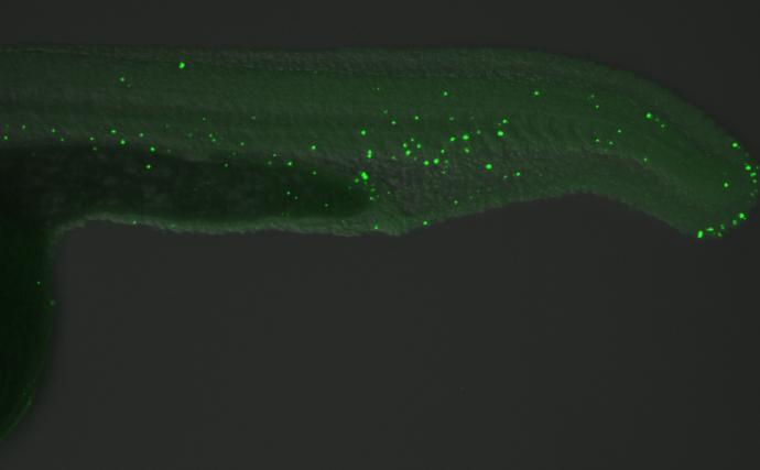
5-Methylcytosine / 5-mC antibody [GT4111] detects 5-Methylcytosine / 5-mC protein on zebrafish by whole mount immunohistochemical analysis. Sample: 24 hours-post-fertilization zebrafish embryo. 5-Methylcytosine / 5-mC antibody [GT4111] dilution: 1:50.Application
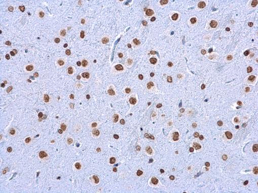
5-Methylcytosine / 5-mC antibody [GT4111] detects 5-Methylcytosine / 5-mC protein at nucleolus in rat brain by immunohistochemical analysis. Sample: Paraffin-embedded rat brain. 5-Methylcytosine / 5-mC antibody [GT4111] diluted at 1:200.Application
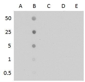
5-methylcytosine antibody [GT4111] Dot blot analysis of anti-5-mC antibody with the synthetic DNA controls . DNA samples (0.5 to 50 ng) were spotted onto positively charged nylon membrane and blotted with 5-mC antibody at a dilution of 1:500. A: Unmethylated DNA fragment B: DNA fragment containing 5-methylcytosine C: DNA fragment containing 5-hydroxymethylcytosine D: DNA fragment containing 5-formylcytosine E: DNA fragment containing 5-carboxylcytosineApplication
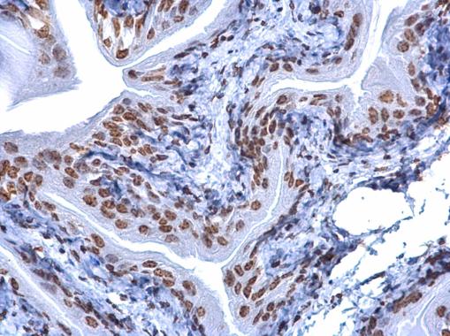
5-Methylcytosine / 5-mC antibody [GT4111] detects 5-Methylcytosine / 5-mC protein at nucleus on mouse urinary bladder by immunohistochemical analysis. Sample: Paraffin-embedded mouse urinary bladder. 5-Methylcytosine / 5-mC antibody [GT4111] dilution: 1:200.Application
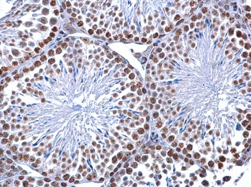
5-Methylcytosine / 5-mC antibody [GT4111] detects 5-Methylcytosine / 5-mC protein at nucleus on mouse testis by immunohistochemical analysis. Sample: Paraffin-embedded mouse testis. 5-Methylcytosine / 5-mC antibody [GT4111] dilution: 1:200.Application
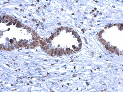
5-Methylcytosine / 5-mC antibody [GT4111] detects 5-Methylcytosine / 5-mC protein at nucleus on human ovarian carcinoma by immunohistochemical analysis. Sample: Paraffin-embedded human ovarian carcinoma. 5-Methylcytosine / 5-mC antibody [GT4111] dilution: 1:200.Application
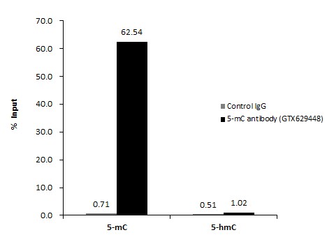
5-Methylcytosine/ 5-mC antibody [GT4111] in MeDIP experiment. Human genomic DNA (500 ng) mixed with methylated cytosine standard kit were subjected to MeDIP with 5-Methylcytosine/ 5-mC antibody [GT4111] at dilution of 1:500 and analyzed with semi-quantitative PCR. The immunoprecipitated DNA was plotted as % of input DNA.| Product Name | 5-Methylcytosine / 5-mC antibody |
|---|---|
| Antibody Type | Primary Antibodies |
| Product description | Mouse Monoclonal antibody [GT4111] to 5-Methylcytosine |
| Clonality | Monoclonal |
|---|---|
| Isotype | IgG1 |
| Clone Number | GT4111 |
| Host Species | Mouse |
| Tested Applications | DotIHC-PIHC-WMeDIP |
| *Optimal dilutions/concentrations should be determined by the researcher.
: |
|
| Concentration | 1mg/ml (Please refer to the vial label for the specific concentration) |
| Purification | Affinity purified |
| Target | 5-Methylcytosine |
|---|
Application
5-Methylcytosine / 5-mC antibody [GT4111] detects 5-Methylcytosine / 5-mC protein on zebrafish by whole mount immunohistochemical analysis. Sample: 24 hours-post-fertilization zebrafish embryo. 5-Methylcytosine / 5-mC antibody [GT4111] dilution: 1:50.
Application
5-Methylcytosine / 5-mC antibody [GT4111] detects 5-Methylcytosine / 5-mC protein at nucleolus in rat brain by immunohistochemical analysis. Sample: Paraffin-embedded rat brain. 5-Methylcytosine / 5-mC antibody [GT4111] diluted at 1:200.
Application
5-methylcytosine antibody [GT4111] Dot blot analysis of anti-5-mC antibody with the synthetic DNA controls . DNA samples (0.5 to 50 ng) were spotted onto positively charged nylon membrane and blotted with 5-mC antibody at a dilution of 1:500. A: Unmethylated DNA fragment B: DNA fragment containing 5-methylcytosine C: DNA fragment containing 5-hydroxymethylcytosine D: DNA fragment containing 5-formylcytosine E: DNA fragment containing 5-carboxylcytosine
Application
5-Methylcytosine / 5-mC antibody [GT4111] detects 5-Methylcytosine / 5-mC protein at nucleus on mouse urinary bladder by immunohistochemical analysis. Sample: Paraffin-embedded mouse urinary bladder. 5-Methylcytosine / 5-mC antibody [GT4111] dilution: 1:200.
Application
5-Methylcytosine / 5-mC antibody [GT4111] detects 5-Methylcytosine / 5-mC protein at nucleus on mouse testis by immunohistochemical analysis. Sample: Paraffin-embedded mouse testis. 5-Methylcytosine / 5-mC antibody [GT4111] dilution: 1:200.
Application
5-Methylcytosine / 5-mC antibody [GT4111] detects 5-Methylcytosine / 5-mC protein at nucleus on human ovarian carcinoma by immunohistochemical analysis. Sample: Paraffin-embedded human ovarian carcinoma. 5-Methylcytosine / 5-mC antibody [GT4111] dilution: 1:200.
Application
5-Methylcytosine/ 5-mC antibody [GT4111] in MeDIP experiment. Human genomic DNA (500 ng) mixed with methylated cytosine standard kit were subjected to MeDIP with 5-Methylcytosine/ 5-mC antibody [GT4111] at dilution of 1:500 and analyzed with semi-quantitative PCR. The immunoprecipitated DNA was plotted as % of input DNA.| Positive Control | GTX400004 , *SH-SY5Y genomic DNA |
|---|---|
| Application Notes | *Optimal dilutions/concentrations should be determined by the researcher.
: |
| Form | Liquid |
|---|---|
| Storage Instructions | Keep as concentrated solution. Aliquot and store at -20ºC or below. Avoid multiple freeze-thaw cycles. |
| Storage Buffer | PBS |


