Application
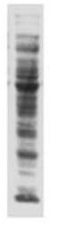
Western blot of acetylated-lysine in HeLA cells usingApplication
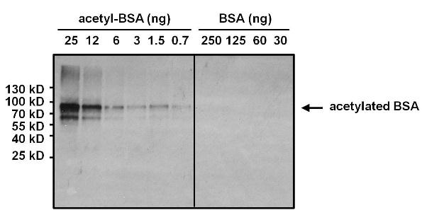
Western blot analysis of Acetyl Lysine was performed by loading various amounts of acetylated BSA or non-acetylated BSA per well and 10ul of PageRuler Prestained Protein Ladder onto a 4-20% Tris-HCl polyacrylamide gel. Proteins were transferred to a PVDF membrane and blocked with 5% BSA/TBST for at least 1 hour. The membrane was probed with an Acetyl Lysine monoclonal antibody at a dilution of 1:1000 overnight at 4 degrees.Application
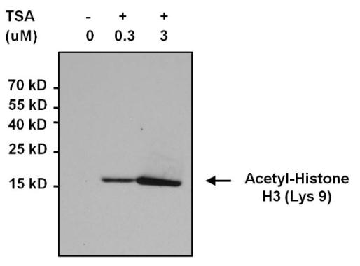
Immunoprecipitation of acetylated Histone H3 (Lys9) was performed using whole cell lysates from cells left untreated (DMSO only) or cells treated with 0.3uM or 3uM Trichostatin A (TSA) for 16 hours. Antigen-antibody complexes were formed by incubating 500ug of the indicated lysate with 3ug of an Acetyl Lysine monoclonal antibody overnight on a rocking platform at 4 degrees.The immune complexes were captured on 50ul Protein A/G Agarose, washed extensively, and eluted with 5X Lane Marker Reducing Sample Buffer. Samples were resolved on a 4-20% Tris-HCl polyacrylamide gel, transferred to a PVDF membrane, and blocked with 5% BSA/TBS-0.1%Tween for at least 1 hour.Application
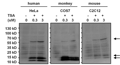
Western blot analysis of lysine acetylated proteins from cells left untreated (DMSO only) or cells treated with 0.3uM or 3uM of Trichostatin A (TSA) for 16 hours was performed by loading 50 ug of the indicated whole cell lysates per well and 10ul of protein ladder onto a 4-20% Tris-HCl polyacrylamide gel. Proteins were transferred to a PVDF membrane and blocked with 5% BSA/TBST for at least 1 hour. The membrane was probed with an Acetyl Lysine monoclonal antibody at a dilution of 1:1000 overnight at 4°C on a rocking platform, washed in TBS-0.1%Tween-20, and probed with a goat anti-mouse IgG-HRP secondary antibody at a dilution of 1:20,000 for 1 hour. Chemiluminescent detection was performed.Application
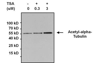
Immunoprecipitation of acetylated alpha-Tubulin was performed using whole cell lysates from cells left untreated (DMSO only) or cells treated with 0.3uM or 3uM Trichostatin A (TSA) for 16 hours. Antigen-antibody complexes were formed by incubating 500ug of lysate with 3ug of an Acetyl Lysine monoclonal antibody overnight on a rocking platform at 4°C. The immune complexes were captured on 50ul Protein A/G Agarose, washed extensively, and eluted. Samples were resolved on a 4-20% Tris-HCl polyacrylamide gel, transferred to a PVDF membrane, and blocked with 5% BSA/TBS-0.1%Tween for at least 1 hour. The membrane was probed with an alpha-Tubulin polyclonal antibody at a dilution of 1:1000 overnight rotating at 4°C, washed in TBST, and probed with Clean-blot IP Detection Reagent at a dilution of 1:2000 for at least 1 hour. Chemiluminescent detection was performedApplication
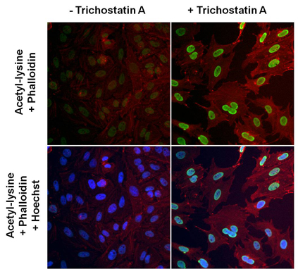
Immunofluorescent analysis of lysine acetylated proteins (green) in HeLa cells left untreated (left panels) or treated with 0.3uM Trichostatin A for 16 hours (right panels). Formalin fixed cells were permeabilized with 0.1% Triton X-100 in TBS for 10 minutes at room temperature and blocked with 1% Blocker BSA) for 15 minutes at room temperature. Cells were probed with an Acetyl Lysine monoclonal antibody at a dilution of 1:100 for 1 hour at room temperature, washed with PBS, and incubated with DyLight 488 goat anti-mouse IgG secondary antibody) at a dilution of 1:400 for 30 minutes at room temperature. F-Actin (red) was stained with DyLight 554 Phalloidin. A merged image with nuclei staining (blue) using Hoechst 33342 dye is shown on bottom panels.Application
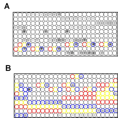
Specific detection of lysine acetylated histones using an Acetyl Lysine monoclonal antibody . Histone peptides containing 384 different modification combinations spotted in duplicate on a peptide array were used according to the manufacturer’s instructions (Active Motif). A schematic and table with representative results (positive detection in red, negative in blue) are shown. Result interpretation and quantification were performed using the manufacturer’s software. Chemiluminescent detection was performed| Product Name | Acetyl Lysine antibody |
|---|---|
| Antibody Type | Primary Antibodies |
| Product description | Mouse Monoclonal antibody to Acetyl LysineIn the nucleus, DNA is tightly packed into nucleosomes generating an environment which is highly repressive towards DNA processes such as transcription. Acetylation of lysine residues within proteins has emerged as an important mechanism used by cells to overcome this repression. The acetylation of non-histone proteins such as transcription factors, as well as histones appears to be involved in this process. Acetylation may result in structural transitions as well as specific signaling within discrete chromatin domains. The role of acetylation in intracellular signaling has been inferred from the binding of acetylated peptides by the conserved bromodomain. Furthermore, recent findings suggest that bromodomain/acetylated-lysine recognition can serve as a regulatory mechanism in protein-protein interactions in numerous cellular processes such as chromatin remodeling and transcriptional activation. |
| Immunogen | Synthetic peptide sequence surrounding the acetylated lysine 9 of histone H3. |
| Clonality | Monoclonal |
|---|---|
| Isotype | IgG |
| Clone Number | 1C6 |
| Host Species | Mouse |
| Tested Applications | DotICC/IFIPWBChIP |
| ICC/IF: 1:100 - 1:500: |
|
| Species Reactivity | HumanChemical |
| Concentration | 1mg/ml (Please refer to the vial label for the specific concentration) |
| Purification | Affinity purified |
| Gene Synonyms | Acetyl Lysine antibody [1C6] Acetyl Lysine |
|---|---|
| Target | Acetyl Lysine |
Application
Western blot of acetylated-lysine in HeLA cells using
Application
Western blot analysis of Acetyl Lysine was performed by loading various amounts of acetylated BSA or non-acetylated BSA per well and 10ul of PageRuler Prestained Protein Ladder onto a 4-20% Tris-HCl polyacrylamide gel. Proteins were transferred to a PVDF membrane and blocked with 5% BSA/TBST for at least 1 hour. The membrane was probed with an Acetyl Lysine monoclonal antibody at a dilution of 1:1000 overnight at 4 degrees.
Application
Immunoprecipitation of acetylated Histone H3 (Lys9) was performed using whole cell lysates from cells left untreated (DMSO only) or cells treated with 0.3uM or 3uM Trichostatin A (TSA) for 16 hours. Antigen-antibody complexes were formed by incubating 500ug of the indicated lysate with 3ug of an Acetyl Lysine monoclonal antibody overnight on a rocking platform at 4 degrees.The immune complexes were captured on 50ul Protein A/G Agarose, washed extensively, and eluted with 5X Lane Marker Reducing Sample Buffer. Samples were resolved on a 4-20% Tris-HCl polyacrylamide gel, transferred to a PVDF membrane, and blocked with 5% BSA/TBS-0.1%Tween for at least 1 hour.
Application
Western blot analysis of lysine acetylated proteins from cells left untreated (DMSO only) or cells treated with 0.3uM or 3uM of Trichostatin A (TSA) for 16 hours was performed by loading 50 ug of the indicated whole cell lysates per well and 10ul of protein ladder onto a 4-20% Tris-HCl polyacrylamide gel. Proteins were transferred to a PVDF membrane and blocked with 5% BSA/TBST for at least 1 hour. The membrane was probed with an Acetyl Lysine monoclonal antibody at a dilution of 1:1000 overnight at 4°C on a rocking platform, washed in TBS-0.1%Tween-20, and probed with a goat anti-mouse IgG-HRP secondary antibody at a dilution of 1:20,000 for 1 hour. Chemiluminescent detection was performed.
Application
Immunoprecipitation of acetylated alpha-Tubulin was performed using whole cell lysates from cells left untreated (DMSO only) or cells treated with 0.3uM or 3uM Trichostatin A (TSA) for 16 hours. Antigen-antibody complexes were formed by incubating 500ug of lysate with 3ug of an Acetyl Lysine monoclonal antibody overnight on a rocking platform at 4°C. The immune complexes were captured on 50ul Protein A/G Agarose, washed extensively, and eluted. Samples were resolved on a 4-20% Tris-HCl polyacrylamide gel, transferred to a PVDF membrane, and blocked with 5% BSA/TBS-0.1%Tween for at least 1 hour. The membrane was probed with an alpha-Tubulin polyclonal antibody at a dilution of 1:1000 overnight rotating at 4°C, washed in TBST, and probed with Clean-blot IP Detection Reagent at a dilution of 1:2000 for at least 1 hour. Chemiluminescent detection was performed
Application
Immunofluorescent analysis of lysine acetylated proteins (green) in HeLa cells left untreated (left panels) or treated with 0.3uM Trichostatin A for 16 hours (right panels). Formalin fixed cells were permeabilized with 0.1% Triton X-100 in TBS for 10 minutes at room temperature and blocked with 1% Blocker BSA) for 15 minutes at room temperature. Cells were probed with an Acetyl Lysine monoclonal antibody at a dilution of 1:100 for 1 hour at room temperature, washed with PBS, and incubated with DyLight 488 goat anti-mouse IgG secondary antibody) at a dilution of 1:400 for 30 minutes at room temperature. F-Actin (red) was stained with DyLight 554 Phalloidin. A merged image with nuclei staining (blue) using Hoechst 33342 dye is shown on bottom panels.
Application
Specific detection of lysine acetylated histones using an Acetyl Lysine monoclonal antibody . Histone peptides containing 384 different modification combinations spotted in duplicate on a peptide array were used according to the manufacturer’s instructions (Active Motif). A schematic and table with representative results (positive detection in red, negative in blue) are shown. Result interpretation and quantification were performed using the manufacturer’s software. Chemiluminescent detection was performed| Application Notes | ICC/IF: 1:100 - 1:500: |
|---|
| Form | Liquid |
|---|---|
| Storage Instructions | Keep as concentrated solution. Aliquot and store at -20ºC or below. Avoid multiple freeze-thaw cycles. |
| Storage Buffer | PBS containing 1mg/ml BSA and 0.05% sodium azide. |


