Application
Adenovirus Type 5 E1A Ab-1 Mouse Monoclonal Antibody, 200µg/mL, BSA and azide| Product Name | Adenovirus type 5 E1A antibody |
|---|---|
| Antibody Type | Primary Antibodies |
| Product description | Mouse monoclonal [M58] to Adenovirus Type 5The early region (E1A) of the adenovirus genome plays a central role in cellular transformation and regulation of gene expression. The E1A region encodes a series of related proteins (35 - 46kDa) with multifunctional capabilities and form a specific complex multifunctional capabilities and form a specific complex with the retinoblastoma tumor suppressor gene product (Rb protein). The E1A and E1B regions together comprise the transforming region of adenovirus. While expression of E1A alone is sufficient to immortalize primary cells, complete transformation requires the additional expression of the E1B region. Several conserved regions of E1A are similar to portions of other viral oncoproteins like the HPV-16 and HPV-18 E7 andSV40 large T antigen. |
| Immunogen | Adenovirus. |
| Clonality | Monoclonal |
|---|---|
| Isotype | IgG2a |
| Clone Number | M58 |
| Host Species | Mouse |
| Tested Applications | ICC/IFIHC-PIPWB |
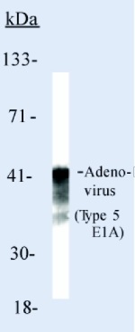
| Immunofluorescence: use at a concentration of 0.2-0.4µg/ml. Immunoprecipitation (Native verified): use at a concentration of 2µg / mg. Western Blotting: use at a concentration of 0.5 - 1.0µg / ml for 2 hrs at RT. Immunohistochemistry (formalin fixed paraffin embedded): use at a concentration of 2 - 4µg / ml for 30 min at RT. Optimal dilutions / concentrations should be determined by the end user. By Western blot, this antibody detects a band of 35-46 kDa, which corresponds to the predicted molecular weight of Adenovirus Type 5. Staining of formalin-fixed tissues requires boiling tissue sections in 10mM citrate buffer, pH 6.0, for 10-20 min followed by cooling at RT for 20 m: |
|
| Species Reactivity | Adenovirus |
| Concentration | 0.2mg/ml |
| Purification | Protein A |
| Gene Synonyms | Adenovirus type5 |
|---|---|
| Target | Adenovirus type 5 |
| Light Chain Type | kappa |
Application
Adenovirus Type 5 E1A Ab-1 Mouse Monoclonal Antibody, 200µg/mL, BSA and azide| Positive Control | 293 cells or adenovirus infected cells and tissues. |
|---|---|
| Application Notes | Immunofluorescence: use at a concentration of 0.2-0.4µg/ml. Immunoprecipitation (Native verified): use at a concentration of 2µg / mg. Western Blotting: use at a concentration of 0.5 - 1.0µg / ml for 2 hrs at RT. Immunohistochemistry (formalin fixed paraffin embedded): use at a concentration of 2 - 4µg / ml for 30 min at RT. Optimal dilutions / concentrations should be determined by the end user. By Western blot, this antibody detects a band of 35-46 kDa, which corresponds to the predicted molecular weight of Adenovirus Type 5. Staining of formalin-fixed tissues requires boiling tissue sections in 10mM citrate buffer, pH 6.0, for 10-20 min followed by cooling at RT for 20 m: |
| Form | Liquid |
|---|---|
| Storage Instructions | Keep as concentrated solution, aliquot and store at 4°C. Do not freeze. |
| Storage Buffer | 10mM PBS, pH7.4, 0.2%BSA, 0.09% sodium azide |


