Application
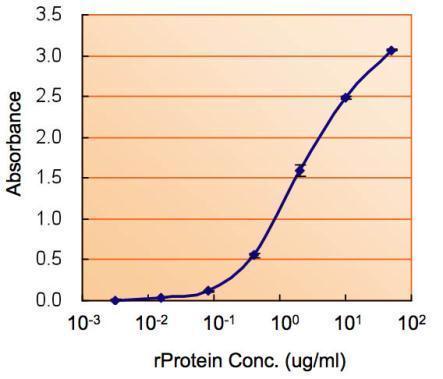
ELISA detection of ALDH2 using for capture at a concentration of 2.5 µg/mL and for detection at a concentration of 1.5 µg/mL.Application
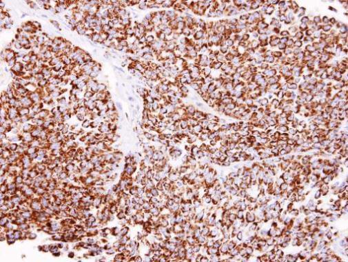
Immunohistochemical analysis of paraffin-embedded AGS xenograft, using ALDH2 antibody at 1:100 dilution.Application
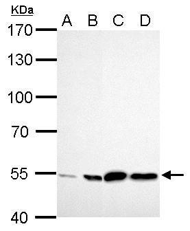
ALDH2 antibody detects ALDH2 protein by western blot analysis. A. 30 µg GL261 whole cell lysate/extract B. 30 µg C8D30 whole cell lysate/extract C. 30 µg BCL-1 whole cell lysae/extract D. 30 µg Raw264.7 whole cell lysate/extract 7.5% SDS-PAGE ALDH2 antibody dilution: 1:1000 The HRP-conjugated anti-rabbit IgG antibody was used to detect the primary antibody.Application
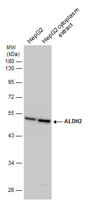
HepG2 whole cell and cytoplasm extracts (30 µg) were separated by 10% SDS-PAGE, and the membrane was blotted with ALDH2 antibody diluted at 1:500. The HRP-conjugated anti-rabbit IgG antibody was used to detect the primary antibody.Application
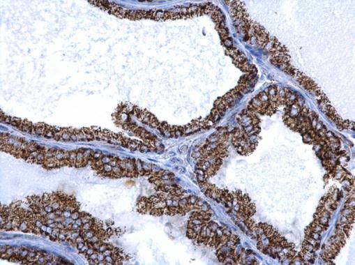
ALDH2 antibody detects ALDH2 protein at mitochondria on mouse prostate by immunohistochemical analysis. Sample: Paraffin-embedded mouse prostate. ALDH2 antibody dilution: 1:500.Application
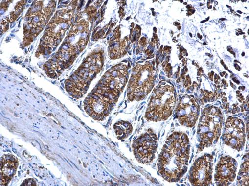
ALDH2 antibody detects ALDH2 protein at mitochondria on mouse colon by immunohistochemical analysis. Sample: Paraffin-embedded mouse colon. ALDH2 antibody dilution: 1:500.Application
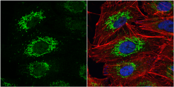
ALDH2 antibody detects ALDH2 protein at mitochondria by immunofluorescent analysis. Sample: A549 cells were fixed in 4% paraformaldehyde at RT for 15 min. Green: ALDH2 protein stained by ALDH2 antibody diluted at 1:500. Red: phalloidin, a cytoskeleton marker, stained by phalloidin (invitrogen, A12380) diluted at 1:200. Blue: Hoechst 33342 staining.| Product Name | ALDH2 antibody |
|---|---|
| Antibody Type | Primary Antibodies |
| Product description | Rabbit Polyclonal antibody to ALDH2 (aldehyde dehydrogenase 2 family (mitochondrial))This protein belongs to the aldehyde dehydrogenase family of proteins. Aldehyde dehydrogenase is the second enzyme of the major oxidative pathway of alcohol metabolism. Two major liver isoforms of this enzyme, cytosolic and mitochondrial, can be distinguished by their electrophoretic mobilities, kinetic properties, and subcellular localizations. Most Caucasians have two major isozymes, while approximately 50% of Orientals have only the cytosolic isozyme, missing the mitochondrial isozyme. A remarkably higher frequency of acute alcohol intoxication among Orientals than among Caucasians could be related to the absence of the mitochondrial isozyme. This gene encodes a mitochondrial isoform, which has a low Km for acetaldehydes, and is localized in mitochondrial matrix. [provided by RefSeq] |
| Immunogen | Recombinant protein encompassing a sequence within the center region of human ALDH2. The exact sequence is proprietary. |
| Clonality | Polyclonal |
|---|---|
| Isotype | IgG |
| Host Species | Rabbit |
| Tested Applications | ELISAICC/IFIHC-PWB |
| *Optimal dilutions/concentrations should be determined by the researcher.
: |
|
| Species Reactivity | HumanMouse |
| Predicted Reactivity | Rat, Xenopus laevis, Pig, BovineRat (96%), Xenopus laevis (82%), Pig (94%), Bovine (94%) |
| Concentration | 0.2mg/ml |
| Purification | Affinity purified |
| Gene Synonyms | ALDH-E2 antibody ALDHI antibody ALDM antibody MGC1806 antibody ALDH2 antibody nucleus-encoded mitochondrial aldehyde dehydrogenase 2 antibody liver mitochondrial ALDH antibody aldehyde dehydrogenase mitochondrial antibody acetaldehyde dehydrogenas |
|---|---|
| Molecular Weight(MW) | 56 kDa (note) (2) |
| Cellular Localization | Mitochondrion matrix |
Application
ELISA detection of ALDH2 using for capture at a concentration of 2.5 µg/mL and for detection at a concentration of 1.5 µg/mL.
Application
Immunohistochemical analysis of paraffin-embedded AGS xenograft, using ALDH2 antibody at 1:100 dilution.
Application
ALDH2 antibody detects ALDH2 protein by western blot analysis. A. 30 µg GL261 whole cell lysate/extract B. 30 µg C8D30 whole cell lysate/extract C. 30 µg BCL-1 whole cell lysae/extract D. 30 µg Raw264.7 whole cell lysate/extract 7.5% SDS-PAGE ALDH2 antibody dilution: 1:1000 The HRP-conjugated anti-rabbit IgG antibody was used to detect the primary antibody.
Application
HepG2 whole cell and cytoplasm extracts (30 µg) were separated by 10% SDS-PAGE, and the membrane was blotted with ALDH2 antibody diluted at 1:500. The HRP-conjugated anti-rabbit IgG antibody was used to detect the primary antibody.
Application
ALDH2 antibody detects ALDH2 protein at mitochondria on mouse prostate by immunohistochemical analysis. Sample: Paraffin-embedded mouse prostate. ALDH2 antibody dilution: 1:500.
Application
ALDH2 antibody detects ALDH2 protein at mitochondria on mouse colon by immunohistochemical analysis. Sample: Paraffin-embedded mouse colon. ALDH2 antibody dilution: 1:500.
Application
ALDH2 antibody detects ALDH2 protein at mitochondria by immunofluorescent analysis. Sample: A549 cells were fixed in 4% paraformaldehyde at RT for 15 min. Green: ALDH2 protein stained by ALDH2 antibody diluted at 1:500. Red: phalloidin, a cytoskeleton marker, stained by phalloidin (invitrogen, A12380) diluted at 1:200. Blue: Hoechst 33342 staining.| Positive Control | GL261 , C8D30 , BCL-1 , Raw264.7 , HepG2 , HepG2 cytoplasm extract |
|---|---|
| Application Notes | *Optimal dilutions/concentrations should be determined by the researcher.
: |
| Form | Liquid |
|---|---|
| Storage Instructions | Keep as concentrated solution. Aliquot and store at -20ºC or below. Avoid multiple freeze-thaw cycles. |
| Storage Buffer | 0.1M Tris, 0.1M Glycine, 10% Glycerol (pH7). 0.01% Thimerosal was added as a preservative. |


