
Application
IHC analysis of mouse hippocampus using alpha Tubulin antibody [236-10501] (green). The section was counterstained with dye to visualize neuronal cell bodies (red) and Hoechst 33258 (blue) to stain nuclei.Application
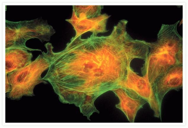
ICC/IF analysis of bovine pulmonary artery endothelial cells using alpha Tubulin antibody [236-10501] (red) and phalloidin (green).Application
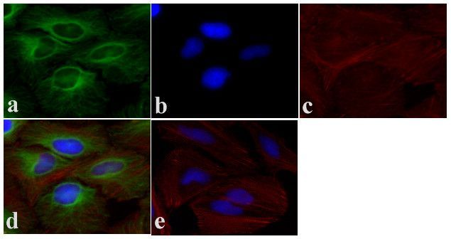
ICC/IF analysis of HeLa cells using alpha Tubulin antibody [236-10501] (green), Phalloidin (red) and DAPI (blue). Panel e shows no primary antibody control.Application
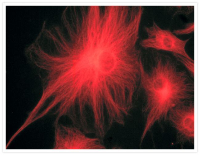
ICC/IF analysis of one bovine pulmonary artery endothelial cell using alpha Tubulin antibody [236-10501].Application
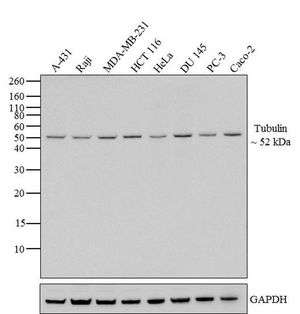
WB analysis of various cell lysates (20 μg per lane) using alpha Tubulin antibody [236-10501] at a dilution of 1:3000.Application
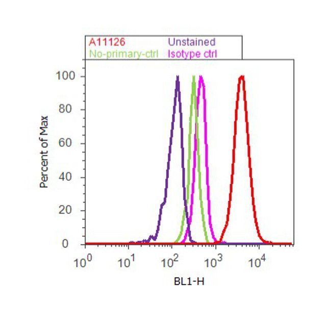
FACS analysis of permeabilized HeLa cells using alpha Tubulin antibody [236-10501] (red) or an isotype control (pink) at a concentration of 3-5 μg/million cells. The purple histogram represents unstained control cells and the green histogram represents no-primary-antibody control.| Product Name | alpha Tubulin antibody [236-10501] |
|---|---|
| Antibody Type | Primary Antibodies |
| Product description | Mouse Monoclonal antibody [236-10501] to alpha Tubulin |
| Immunogen | Bovine Alpha-Tubulin. |
| Clonality | Monoclonal |
|---|---|
| Isotype | IgG1 |
| Clone Number | 236-10501 |
| Host Species | Mouse |
| Tested Applications | FACSICC/IFIHCWB |
| FACS: 1:100 - 1:500. ICC/IF: 1:100 - 1:500. WB: 1:1000 - 1:5000 | |
| Species Reactivity | HumanMouseBovine |
| Concentration | 1mg/ml |
| Gene Synonyms | tubulin, alpha 4a , TUBA4A |
|---|---|
| Target | alpha Tubulin |
Application
IHC analysis of mouse hippocampus using alpha Tubulin antibody [236-10501] (green). The section was counterstained with dye to visualize neuronal cell bodies (red) and Hoechst 33258 (blue) to stain nuclei.
Application
ICC/IF analysis of bovine pulmonary artery endothelial cells using alpha Tubulin antibody [236-10501] (red) and phalloidin (green).
Application
ICC/IF analysis of HeLa cells using alpha Tubulin antibody [236-10501] (green), Phalloidin (red) and DAPI (blue). Panel e shows no primary antibody control.
Application
ICC/IF analysis of one bovine pulmonary artery endothelial cell using alpha Tubulin antibody [236-10501].
Application
WB analysis of various cell lysates (20 μg per lane) using alpha Tubulin antibody [236-10501] at a dilution of 1:3000.
Application
FACS analysis of permeabilized HeLa cells using alpha Tubulin antibody [236-10501] (red) or an isotype control (pink) at a concentration of 3-5 μg/million cells. The purple histogram represents unstained control cells and the green histogram represents no-primary-antibody control.| Application Notes | FACS: 1:100 - 1:500. ICC/IF: 1:100 - 1:500. WB: 1:1000 - 1:5000 |
|---|
| Form | Liquid |
|---|---|
| Storage Instructions | -20°C |
| Storage Buffer | phosphate buffered saline , pH 7.4, 150mM NaCl, 0.02% sodium azide and 50% glycerol. |


