

Application
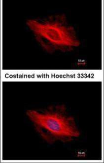
Immunofluorescence analysis of methanol-fixed HeLa, using alpha Tubulin antibody at 1:500 dilution.Application
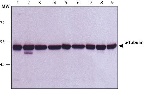
WB analysis of (1) HeLa, (2) Jurkat, (3) COS7, (4) NIH-3T3, (5) PC-12, (6) RAT2, (7) CHO, (8) MDBK and (9) MDCK lysates using alpha Tubulin antibody [B-5-1-2] at a concentration of 1 μg/ml.Application
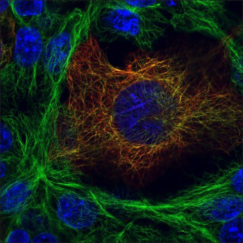
ICC/IF analysis of HEK293T cells transfected with a HA tagged protein using alpha Tubulin antibody [B-5-1-2] (green), HA antibody (red) and DAPI (blue).WB
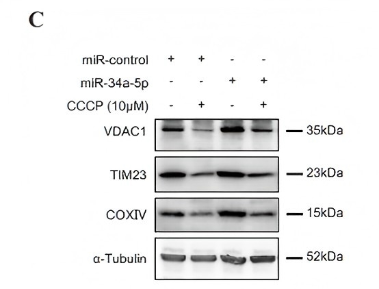
(C) Protein levels of VDAC1, T1M23 and COXIV in HEK293 cells were immunoblotted and quantified. After transfection of miR-34a-5p for 48 h, HEK293 cells were treated with CCCP for 24 h.WB
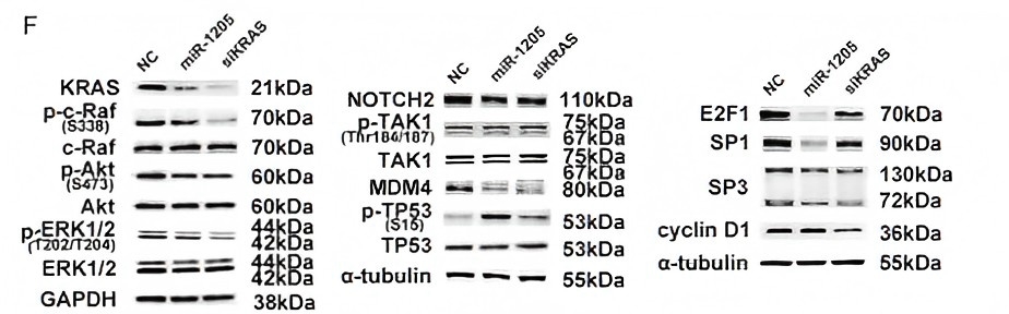
F. QRT-PCR and WB analysis of genes which both were predicted to be targeted by miR-1205 in the KRAS signaling pathway.WB
I. A549 or H460 cells were transfected with different dose of siKRAS (5, 10 nM) or NC for 72 h, p-ERK1/2, ERK1/2 and MDM4 protein levels were detected. J. A549 or H460 cells were treated with SCH772984 (0, 0.1, 1, 10 μM) for 24 h, and analyzed for the expression of p-ERK1/2 and MDM4.WB
I. A549 or H460 cells were transfected with different dose of siKRAS (5, 10 nM) or NC for 72 h, KRAS signaling and E2F1 protein level were analyzed. J. A549 or H460 cells were treated with SCH772984 (0, 0.1, 1, 10 μM) for 24 h, and analyzed for the expression of p-ERK1/2 and E2F1.WB
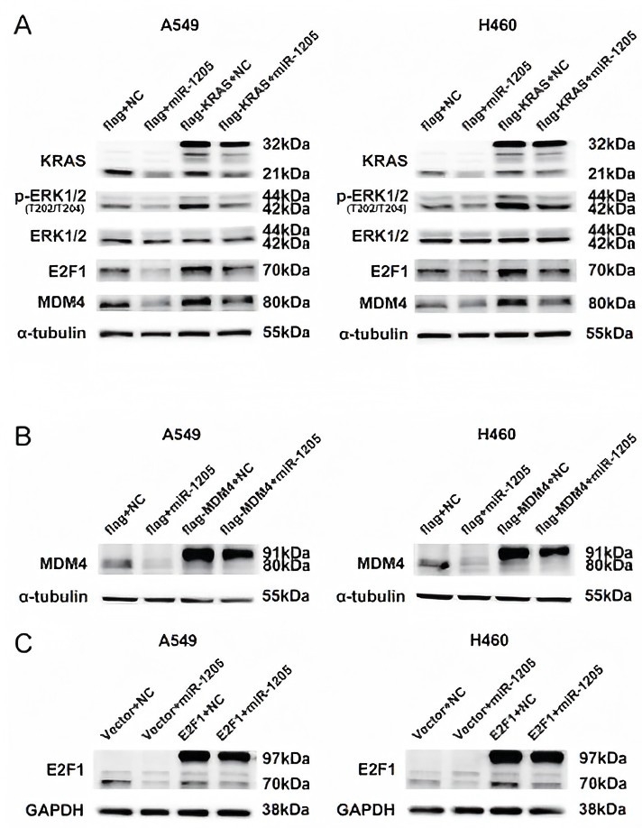
A. p-ERK1/2, ERK1/2, MDM4 and E2F1 protein levels were measured by western blot in A549 and H460 cells after transfected with indicated miRNA mimics or plasmids. B and C. MDM4 and E2F1 protein levels were detected after transfected with indicated miRNA mimics or plasmidsWB
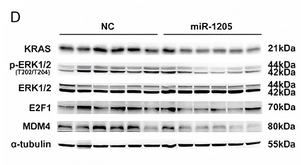
D. KRAS signaling, MDM4 and E2F1 protein levels of NC group and miR-1205 group were detected and analyzed.| Product Name | alpha Tubulin antibody [B-5-1-2] |
|---|---|
| Antibody Type | Primary Antibodies |
| Product description | Mouse monoclonal [B-5-1-2] to alpha TubulinTubulin is the major building block of microtubules. This intracellular cylindrical filamentous structure is present in almost all eukaryotic cells. Microtubules function as structural and mobile elements in mitosis, intracellular transport, flagellar movement and in the cytoskeleton. Tubulin is a heterodimer which consists of alpha tubulin and beta tubulin. |
| Immunogen | Sarkosyl-resistant filaments from Stronglycentrotus purpuratus (sea urchin). |
| Clonality | Monoclonal |
|---|---|
| Isotype | IgG1 |
| Clone Number | B-5-1-2 |
| Host Species | Mouse |
| Tested Applications | IBICC/IFIPRIAWB |
| WB:1:500~1:2000 IP:1:100~1:500 ICC/IF:1:100~1:500 |
|
| Species Reactivity | BovineChlamydomonas ReinhardtiiHumanKangarooMonkeyMouseRatSea Urchin |
| Concentration | 1mg/ |
| Purification | Protein A |
| Gene Synonyms | TUBA1, P68366, alpha Tubulin, 191110, H2ALPHA, H2-ALPHA, 7277, FLJ30169, H2 ALPHA |
|---|---|
| Target | alpha Tubulin |
| Molecular Weight(MW) | 50 kDa |
| Cellular Localization | Major constituent of microtubules. |
Application
Immunofluorescence analysis of methanol-fixed HeLa, using alpha Tubulin antibody at 1:500 dilution.
Application
WB analysis of (1) HeLa, (2) Jurkat, (3) COS7, (4) NIH-3T3, (5) PC-12, (6) RAT2, (7) CHO, (8) MDBK and (9) MDCK lysates using alpha Tubulin antibody [B-5-1-2] at a concentration of 1 μg/ml.
Application
ICC/IF analysis of HEK293T cells transfected with a HA tagged protein using alpha Tubulin antibody [B-5-1-2] (green), HA antibody (red) and DAPI (blue).
WB
(C) Protein levels of VDAC1, T1M23 and COXIV in HEK293 cells were immunoblotted and quantified. After transfection of miR-34a-5p for 48 h, HEK293 cells were treated with CCCP for 24 h.
WB
F. QRT-PCR and WB analysis of genes which both were predicted to be targeted by miR-1205 in the KRAS signaling pathway.
WB
I. A549 or H460 cells were transfected with different dose of siKRAS (5, 10 nM) or NC for 72 h, p-ERK1/2, ERK1/2 and MDM4 protein levels were detected. J. A549 or H460 cells were treated with SCH772984 (0, 0.1, 1, 10 μM) for 24 h, and analyzed for the expression of p-ERK1/2 and MDM4.
WB
I. A549 or H460 cells were transfected with different dose of siKRAS (5, 10 nM) or NC for 72 h, KRAS signaling and E2F1 protein level were analyzed. J. A549 or H460 cells were treated with SCH772984 (0, 0.1, 1, 10 μM) for 24 h, and analyzed for the expression of p-ERK1/2 and E2F1.
WB
A. p-ERK1/2, ERK1/2, MDM4 and E2F1 protein levels were measured by western blot in A549 and H460 cells after transfected with indicated miRNA mimics or plasmids. B and C. MDM4 and E2F1 protein levels were detected after transfected with indicated miRNA mimics or plasmids
WB
D. KRAS signaling, MDM4 and E2F1 protein levels of NC group and miR-1205 group were detected and analyzed.| Positive Control | Total cell extract of the human foreskin fibroblast cell line (FS11) |
|---|---|
| Application Notes | WB:1:500~1:2000 IP:1:100~1:500 ICC/IF:1:100~1:500 |
| Form | Liquid |
|---|---|
| Storage Instructions | Aliquot and store at -20°C or -80°C. Avoid repeated freeze / thaw cycles. |
| Storage Buffer | Preservative: 15mM Sodium Azide; Constituents: 0.01M PBS. pH 7.4 |


