Application
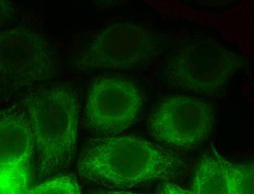
Immunofluorescence analysis of paraformaldehyde-fixed Hela, using alpha Tubulin antibody [DM1A] at 1:1000 dilution.Application
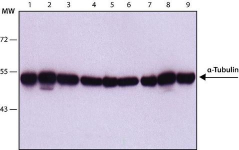
WB analysis of (1) HeLa, (2) Jurkat, (3) COS7, (4) NIH-3T3, (5) PC-12, (6) RAT2, (7) CHO, (8) MDBK and (9) MDCK lysates using alpha Tubulin antibody [DM1A] at a concentration of 1 μg/ml.Application
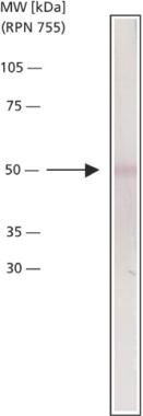
Chicken gizzard extract was separated on SDS-PAGE and probed with Monoclonal PA to Alpha Tubulin (Cat. No. ). The antibody was developed with Anti-Mouse IgG (Fab specific), Alkaline Phosphatase conjugate ( and a NBT/BCIP substrate. Antibody concentration 1 μg/mLApplication
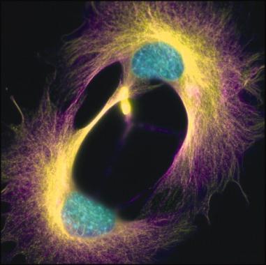
Lamin A/C knockout mice embryo fibroblasts following mitosis were stained using Monoclonal Anti-α-Tubulin, clone no. DM1A (Cat. No. ). Staining patterns show actin (yellow), microtubules (purple) and DNA (cyan).Application
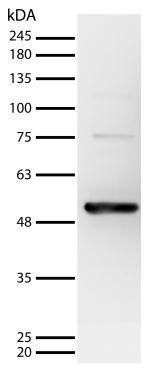
at 1:1000 Sample: Hela whole cell lysate Secondary: Mouse IgG antibody (HRP)| Product Name | alpha Tubulin antibody [DM1A] (purified) |
|---|---|
| Antibody Type | Primary Antibodies |
| Product description | Mouse monoclonal antibody [DM1A] to alpha TubulinTubulin is the major building block of microtubules. This intracellular cylindrical filamentous structure is present in almost all eukaryotic cells. Microtubules function as structural and mobile elements in mitosis, intracellular transport, flagellar movement and in the cytoskeleton. Tubulin is a heterodimer which consists of alpha-tubulin and beta-tubulin; both subunits have a molecular weight of approximately 50 kDa and share considerable homology. The most widely studied tubulins have been isolated from vertebrate brains. The microtubules can be viewed in immunofluorescent microscopy, which enables the observation of the intracellular organization of proteins that are in the form of a supramolecular structure. |
| Immunogen | Native chicken brain microtubules |
| Clonality | Monoclonal |
|---|---|
| Isotype | IgG1 |
| Clone Number | DM1A |
| Host Species | Mouse |
| Tested Applications | EMICC/IFIHC-PIPWBIB |
| Electron Microscopy: Use at an assay dependent dilution. IF: Use at an assay dependent dilution. IHC-P: Use at an assay dependent dilution. Antigen retrieval is not essential but may optimize staining. IP: Use at an assay dependent dilution. WB: Use at a dilution of about 1/5000. Detects a band of approximately 57 kDa. This antibody is ideal for use as a Western blot loading control. Optimal dilutions/concentrations should be determined by the end user. | |
| Species Reactivity | HumanMousePigRatGerbilGuinea PigXenopusBovine |
| Concentration | 1mg/ml (Please refer to the vial label for the specific concentration) |
| Purification | Unpurified |
| Target | alpha Tubulin |
|---|---|
| Cellular Localization | Major constituent of Microtubules |
| Light Chain Type | kappa |
Application
Immunofluorescence analysis of paraformaldehyde-fixed Hela, using alpha Tubulin antibody [DM1A] at 1:1000 dilution.
Application
WB analysis of (1) HeLa, (2) Jurkat, (3) COS7, (4) NIH-3T3, (5) PC-12, (6) RAT2, (7) CHO, (8) MDBK and (9) MDCK lysates using alpha Tubulin antibody [DM1A] at a concentration of 1 μg/ml.
Application
Chicken gizzard extract was separated on SDS-PAGE and probed with Monoclonal PA to Alpha Tubulin (Cat. No. ). The antibody was developed with Anti-Mouse IgG (Fab specific), Alkaline Phosphatase conjugate ( and a NBT/BCIP substrate. Antibody concentration 1 μg/mL
Application
Lamin A/C knockout mice embryo fibroblasts following mitosis were stained using Monoclonal Anti-α-Tubulin, clone no. DM1A (Cat. No. ). Staining patterns show actin (yellow), microtubules (purple) and DNA (cyan).
Application
at 1:1000 Sample: Hela whole cell lysate Secondary: Mouse IgG antibody (HRP)| Application Notes | Electron Microscopy: Use at an assay dependent dilution. IF: Use at an assay dependent dilution. IHC-P: Use at an assay dependent dilution. Antigen retrieval is not essential but may optimize staining. IP: Use at an assay dependent dilution. WB: Use at a dilution of about 1/5000. Detects a band of approximately 57 kDa. This antibody is ideal for use as a Western blot loading control. Optimal dilutions/concentrations should be determined by the end user. |
|---|
| Form | Liquid |
|---|---|
| Storage Instructions | Keep as concentrated solution. Store at 4ºC short term. For extended storage aliquot and store at -20ºC or below. Avoid freeze-thaw cycles. |
| Storage Buffer | Phosphate-buffered saline, containing 0.02% sodium azide |


