Application
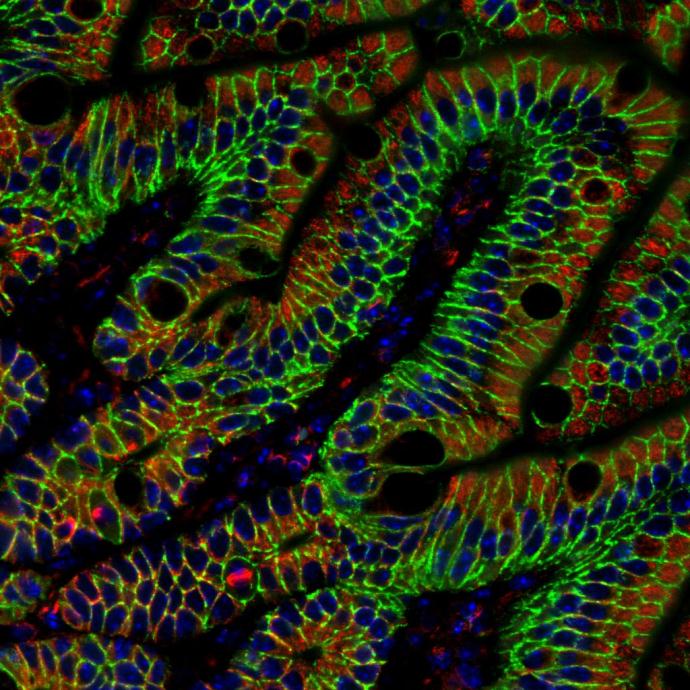
alpha Tubulin antibody detects alpha Tubulin protein at cytoplasm in mouse colon by immunohistochemical analysis. Sample: Paraffin-embedded mouse colon. Red: alpha Tubulin antibody diluted at 1:500. Green: beta Catenin antibody [N1N2-2] diluted at 1:500. Blue: Hoechst 33342 staining.Application
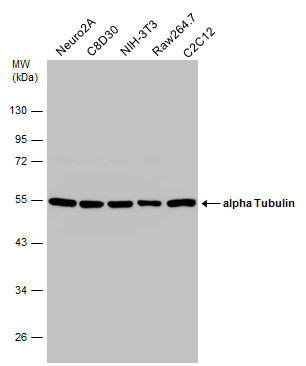
Various whole cell extracts (30 µg) were separated by 10% SDS-PAGE, and the membrane was blotted with alpha Tubulin antibody diluted at 1:1000. The HRP-conjugated anti-mouse IgG antibody was used to detect the primary antibody.Application
TUBA1B antibody detects TUBA1B protein at Cytoskeleton by immunofluorescent analysis. Sample: HeLa cells were fixed in -20℃ 100% methanol for 5 min. Green: TUBA1B protein stained by TUBA1B antibody diluted at 1:500. Blue: Hoechst 33342 staining.Application
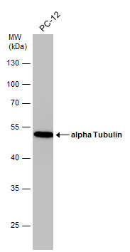
Whole cell extract (30 µg) was separated by 10% SDS-PAGE, and the membrane was blotted with alpha Tubulin antibody diluted at 1:1000. The HRP-conjugated anti-mouse IgG antibody was used to detect the primary antibody.Application
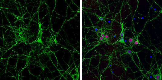
alpha Tubulin antibody detects alpha Tubulin protein expression by immunofluorescent analysis. Sample: Cultured rat E18 primary cortical neuron, DIV 8. Cells were fixed in 4% paraformaldehyde at RT for 15 min. Green: alpha Tubulin protein stained by alpha Tubulin antibody diluted at 1:250. Red: NeuN, stained by NeuN antibody diluted at 1:250. Blue: Fluoroshield with DAPI .Application
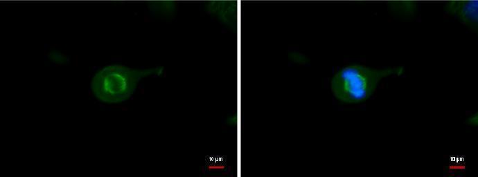
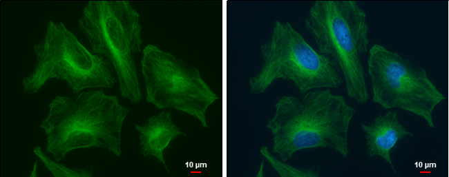
alpha Tubulin antibody detects alpha Tubulin protein at cytoskeleton by immunofluorescent analysis. Sample: HeLa cells were fixed in 4% paraformaldehyde at RT for 15 min. Green: alpha Tubulin protein stained by alpha Tubulin antibody diluted at 1:500. Blue: Hoechst 33342 staining. Scale bar = 10 μm.Application
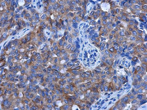
alpha Tubulin antibody detects alpha Tubulin protein at cytoplasm in human lung cancer by immunohistochemical analysis. Sample: Paraffin-embedded human lung cancer. alpha Tubulin antibody [GT114] diluted at 1:500.Application
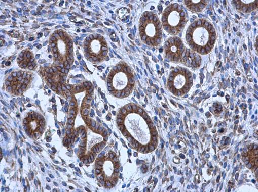
alpha Tubulin antibody detects alpha Tubulin protein at cytoplasm in human cervix by immunohistochemical analysis. Sample: Paraffin-embedded human cervix. alpha Tubulin antibody diluted at 1:500.Application
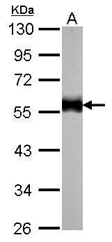
alpha Tubulin antibody detects TUBA1B protein by western blot analysis. A. 30 µg zebrafish eye lysate/extract 10% SDS-PAGE alpha Tubulin antibody dilution: 1:1000 The HRP-conjugated anti-mouse IgG antibody was used to detect the primary antibody.Application
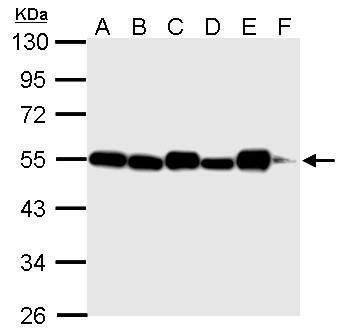
alpha Tubulin antibody detects TUBA1B protein by western blot analysis. A. 30 µg 293T whole cell lysate/extract B. 30 µg NIH-3T3 whole cell lysate/extract C. 30 µg mouse brain lysate/extract D. 30 µg PC-12 whole cell lysate/extract E. 30 µg rat brain lysate/extract F. 30 µg drosophila lysae/extract 10% SDS-PAGE alpha Tubulin antibody dilution: 1:5000 The HRP-conjugated anti-mouse IgG antibody was used to detect the primary antibody.Application
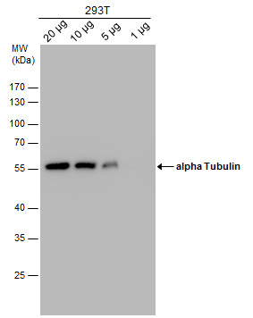
alpha Tubulin antibody detects alpha Tubulin protein by western blot analysis. Varied amount of 293T Whole cell extracts was separated by 10% SDS-PAGE, and the membrane was blotted with alpha Tubulin antibody at a dilution of 1:1000. The HRP-conjugated anti-mouse IgG antibody was used to detect the primary antibody.Application
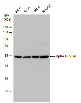
alpha Tubulin antibody detects alpha Tubulin protein by western blot analysis. Various whole cell extracts (30 µg) were separated by 10% SDS-PAGE, and the membrane was blotted with alpha Tubulin antibody diluted at a dilution of 1:1000. The HRP-conjugated anti-mouse IgG antibody was used to detect the primary antibody.Application
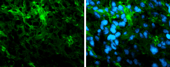
Immunofluorescence photomicrographs of frozen sections of mouse brain. Green: alpha Tubulin antibody diluted at 1:200. The signal was developed using goat anti-rmouse IgG antibody (Dylight488) . Blue: Nuclear staining with Hoechst 33342.| Product Name | alpha Tubulin antibody [GT114] |
|---|---|
| Antibody Type | Primary Antibodies |
| Product description | Mouse Monoclonal antibody [GT114] to alpha Tubulin (tubulin, alpha 1b)Tubulin is the major constituent of microtubules. It binds two moles of GTP, one at an exchangeable site on the beta chain and one at a non-exchangeable site on the alpha-chain. |
| Immunogen | Recombinant protein encompassing a sequence within the center region of human alpha Tubulin. The exact sequence is proprietary. |
| Clonality | Monoclonal |
|---|---|
| Isotype | IgG2a |
| Clone Number | GT114 |
| Host Species | Mouse |
| Tested Applications | ICC/IFIHC-FIHC-PWB |
| WB:1:500~1:1000 ICC/IF:1:100~1:500 IHC:1:50~1:200 *Optimal dilutions/concentrations should be determined by the researcher. |
|
| Species Reactivity | DrosophilaHumanMouseRatZebra Fish |
| Predicted Reactivity | Xenopus laevis, Pig, Rabbit, Rhesus Monkey, Chimpanzee, BovineXenopus laevis (95%), Pig (100%), Rabbit (100%), Rhesus Monkey (100%), Chimpanzee (99%), Bovine (100%) |
| Concentration | 1mg/ml (Please refer to the vial label for the specific concentration) |
| Purification | Affinity purified |
| Gene Synonyms | K-ALPHA-1 antibody, TUBA1B antibody, alpha-tubulin ubiquitous antibody, alpha tubulin antibody, tubulin K-alpha-1 antibody, tubulin alpha-ubiquitous chain antibody, tubulin, alpha, ubiquitous antibody, tubulin alpha-1B chain antibody, tubulin, alpha 1b ant |
|---|---|
| Molecular Weight(MW) | 50 kDa (note) |
Application
alpha Tubulin antibody detects alpha Tubulin protein at cytoplasm in mouse colon by immunohistochemical analysis. Sample: Paraffin-embedded mouse colon. Red: alpha Tubulin antibody diluted at 1:500. Green: beta Catenin antibody [N1N2-2] diluted at 1:500. Blue: Hoechst 33342 staining.
Application
Various whole cell extracts (30 µg) were separated by 10% SDS-PAGE, and the membrane was blotted with alpha Tubulin antibody diluted at 1:1000. The HRP-conjugated anti-mouse IgG antibody was used to detect the primary antibody.
Application
TUBA1B antibody detects TUBA1B protein at Cytoskeleton by immunofluorescent analysis. Sample: HeLa cells were fixed in -20℃ 100% methanol for 5 min. Green: TUBA1B protein stained by TUBA1B antibody diluted at 1:500. Blue: Hoechst 33342 staining.
Application
Whole cell extract (30 µg) was separated by 10% SDS-PAGE, and the membrane was blotted with alpha Tubulin antibody diluted at 1:1000. The HRP-conjugated anti-mouse IgG antibody was used to detect the primary antibody.
Application
alpha Tubulin antibody detects alpha Tubulin protein expression by immunofluorescent analysis. Sample: Cultured rat E18 primary cortical neuron, DIV 8. Cells were fixed in 4% paraformaldehyde at RT for 15 min. Green: alpha Tubulin protein stained by alpha Tubulin antibody diluted at 1:250. Red: NeuN, stained by NeuN antibody diluted at 1:250. Blue: Fluoroshield with DAPI .
Application
alpha Tubulin antibody detects alpha Tubulin protein at cytoskeleton by immunofluorescent analysis. Sample: HeLa cells were fixed in 4% paraformaldehyde at RT for 15 min. Green: alpha Tubulin protein stained by alpha Tubulin antibody diluted at 1:500. Blue: Hoechst 33342 staining. Scale bar = 10 μm.
Application
alpha Tubulin antibody detects alpha Tubulin protein at cytoplasm in human lung cancer by immunohistochemical analysis. Sample: Paraffin-embedded human lung cancer. alpha Tubulin antibody [GT114] diluted at 1:500.
Application
alpha Tubulin antibody detects alpha Tubulin protein at cytoplasm in human cervix by immunohistochemical analysis. Sample: Paraffin-embedded human cervix. alpha Tubulin antibody diluted at 1:500.
Application
alpha Tubulin antibody detects TUBA1B protein by western blot analysis. A. 30 µg zebrafish eye lysate/extract 10% SDS-PAGE alpha Tubulin antibody dilution: 1:1000 The HRP-conjugated anti-mouse IgG antibody was used to detect the primary antibody.
Application
alpha Tubulin antibody detects TUBA1B protein by western blot analysis. A. 30 µg 293T whole cell lysate/extract B. 30 µg NIH-3T3 whole cell lysate/extract C. 30 µg mouse brain lysate/extract D. 30 µg PC-12 whole cell lysate/extract E. 30 µg rat brain lysate/extract F. 30 µg drosophila lysae/extract 10% SDS-PAGE alpha Tubulin antibody dilution: 1:5000 The HRP-conjugated anti-mouse IgG antibody was used to detect the primary antibody.
Application
alpha Tubulin antibody detects alpha Tubulin protein by western blot analysis. Varied amount of 293T Whole cell extracts was separated by 10% SDS-PAGE, and the membrane was blotted with alpha Tubulin antibody at a dilution of 1:1000. The HRP-conjugated anti-mouse IgG antibody was used to detect the primary antibody.
Application
alpha Tubulin antibody detects alpha Tubulin protein by western blot analysis. Various whole cell extracts (30 µg) were separated by 10% SDS-PAGE, and the membrane was blotted with alpha Tubulin antibody diluted at a dilution of 1:1000. The HRP-conjugated anti-mouse IgG antibody was used to detect the primary antibody.
Application
Immunofluorescence photomicrographs of frozen sections of mouse brain. Green: alpha Tubulin antibody diluted at 1:200. The signal was developed using goat anti-rmouse IgG antibody (Dylight488) . Blue: Nuclear staining with Hoechst 33342.| Positive Control | 293T , A431 , HeLa , HepG2 , NIH-3T3 , mouse brain , PC-12 , rat brain , drosophila , zebrafish eye , Neuro2A , C8D30 , NIH-3T3 , Raw264.7 , C2C12 , PC-12 , *U87-MG |
|---|---|
| Application Notes | WB:1:500~1:1000 ICC/IF:1:100~1:500 IHC:1:50~1:200 *Optimal dilutions/concentrations should be determined by the researcher. |
| Form | Liquid |
|---|---|
| Storage Instructions | For short-term storage, store at 4° C. For long-term storage, aliquot and store at -20ºC or below. Avoid multiple freeze-thaw cycles. |
| Storage Buffer | phosphate buffered saline , pH 7.4, 150mM NaCl, 0.02% sodium azide and 50% glycerol. |


