Application
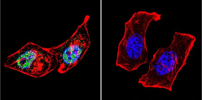
Immunofluorescent analysis of AP2 in HeLa cells. AP2 staining (green), F-Actin staining with Phalloidin (red) and nuclei with DAPI (blue) is shown. Cells were grown on slides and fixed with formaldehyde prior to staining. Cells were probed without (control) or with AP2 antibody [3B5] at a dilution of 1:100 over night at 4?C, washed with PBS and incubated with a proper secondary antibody. Images were taken at 60X magnification.Application
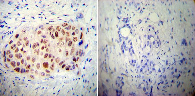
Immunohistochemistry was performed on cancer biopsies of deparaffinized human breast carcinoma tissue. To expose target proteins, heat induced antigen retrieval was performed using 10mM sodium citrate (pH6.0) buffer, microwaved for 8-15 minutes. Following antigen retrieval tissues were blocked in 3% BSA-PBS for 30 minutes at room temperature. Tissues were then probed at a dilution of 1:50 with or without AP2 antibody [3B5] overnight at 4°C in a humidified chamber. Tissues were washed extensively with PBST and endogenous peroxidase activity was quenched with a peroxidase suppressor. Detection was performed using a biotin-conjugated secondary antibody and SA-HRP, followed by colorimetric detection using DAB. Tissues were counterstained with hematoxylin and prepped for mounting.| Product Name | AP2 alpha antibody [3B5] |
|---|---|
| Antibody Type | Primary Antibodies |
| Product description | Mouse monoclonal antibody [3B5] to AP2 alphaThe protein encoded by this gene is a transcription factor that binds the consensus sequence 5'-GCCNNNGGC-3'. The encoded protein functions as either a homodimer or as a heterodimer with similar family members. This protein activates the transcription of some genes while inhibiting the transcription of others. Defects in this gene are a cause of branchiooculofacial syndrome (BOFS). Three transcript variants encoding different isoforms have been found for this gene.[provided by RefSeq, Dec 2009] |
| Immunogen | N-terminus of human AP2 alpha. |
| Clonality | Monoclonal |
|---|---|
| Isotype | IgG2 |
| Clone Number | 3B5 |
| Host Species | Mouse |
| Tested Applications | ICC/IFIHCIHC-PIPWB |
| IHC: 1:50 | |
| Species Reactivity | HumanMouseChicken |
| Concentration | 1mg/ml |
| Purification | Protein G |
| Gene Synonyms | TFAP2 , AP-2 , AP2TF , transcription factor AP-2 alpha (activating enhancer binding protein 2 alpha) , BOFS , TFAP2A , AP-2alpha |
|---|---|
| Target | AP2 alpha |
Application
Immunofluorescent analysis of AP2 in HeLa cells. AP2 staining (green), F-Actin staining with Phalloidin (red) and nuclei with DAPI (blue) is shown. Cells were grown on slides and fixed with formaldehyde prior to staining. Cells were probed without (control) or with AP2 antibody [3B5] at a dilution of 1:100 over night at 4?C, washed with PBS and incubated with a proper secondary antibody. Images were taken at 60X magnification.
Application
Immunohistochemistry was performed on cancer biopsies of deparaffinized human breast carcinoma tissue. To expose target proteins, heat induced antigen retrieval was performed using 10mM sodium citrate (pH6.0) buffer, microwaved for 8-15 minutes. Following antigen retrieval tissues were blocked in 3% BSA-PBS for 30 minutes at room temperature. Tissues were then probed at a dilution of 1:50 with or without AP2 antibody [3B5] overnight at 4°C in a humidified chamber. Tissues were washed extensively with PBST and endogenous peroxidase activity was quenched with a peroxidase suppressor. Detection was performed using a biotin-conjugated secondary antibody and SA-HRP, followed by colorimetric detection using DAB. Tissues were counterstained with hematoxylin and prepped for mounting.| Application Notes | IHC: 1:50 |
|---|
| Form | Liquid |
|---|---|
| Storage Instructions | -20° C, Avoid Freeze/Thaw Cycles |
| Storage Buffer | PBS containing 1 mg/ml BSA and 0.05 % sodium azide |


