Application
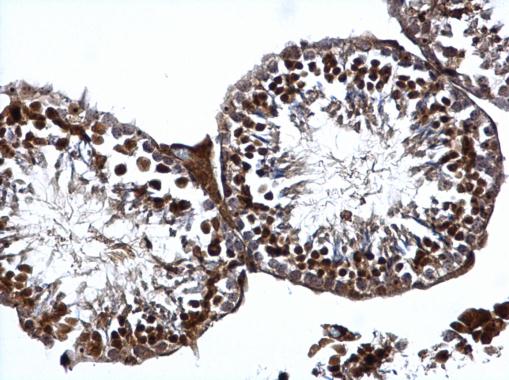
ATR antibody detects ATR protein at nucleus on mouse testis by immunohistochemical analysis. Sample: Paraffin-embedded mouse testis. ATR antibody dilution: 1:500.Application
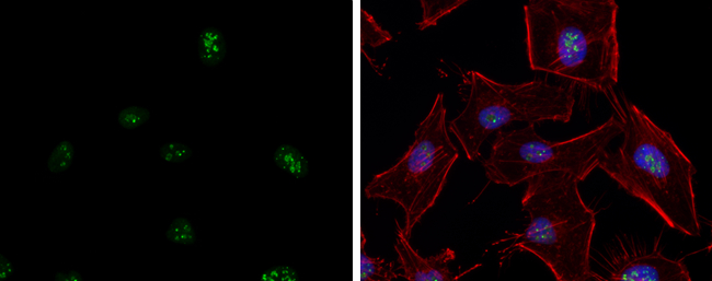
ATR antibody detects ATR protein at nucleolus by immunofluorescent analysis. Sample: HeLa cells were fixed in 4% paraformaldehyde at RT for 15 min. Green: ATR protein stained by ATR antibody diluted at 1:5000. Red: phalloidin, a cytoskeleton marker, diluted at 1:200. Blue: Hoechst 33342 staining. Scale bar = 10 μm.Application
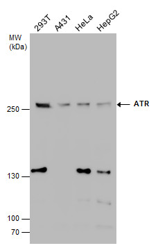
ATR antibody detects ATR protein by western blot analysis. Various whole cell extracts (30 µg) were separated by 5% SDS-PAGE, and the membrane was blotted with ATR antibody diluted at a dilution of 1:500. The HRP-conjugated anti-rabbit IgG antibody was used to detect the primary antibody.Application
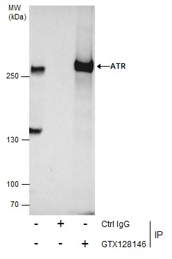
Immunoprecipitation of ATR protein from HCT-116 whole cell extracts treated with Hydroxyurea for 6 hr using 5 μg of ATR antibody . Western blot analysis was performed using ATR antibody . EasyBlot anti-Rabbit IgG was used as a secondary reagent.Application
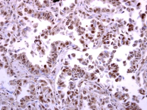
ATR antibody detects ATR protein at nucleus on human lung carcinoma by immunohistochemical analysis. Sample: Paraffin-embedded human lung carcinoma. ATR antibody diluted at 1:250.Application
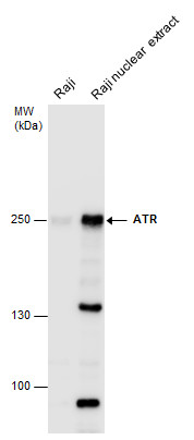
ATR antibody detects ATR protein by western blot analysis. Raji whole cell extracts and nuclear extracts (30 µg) were separated by 5% SDS-PAGE, and the membrane was blotted with ATR antibody at a dilution of 1:500. The HRP-conjugated anti-rabbit IgG antibody was used to detect the primary antibody.| Product Name | ATR antibody |
|---|---|
| Antibody Type | Primary Antibodies |
| Product description | Rabbit Polyclonal antibody to ATR (ataxia telangiectasia and Rad3 related)The protein encoded by this gene belongs the PI3/PI4-kinase family, and is most closely related to ATM, a protein kinase encoded by the gene mutated in ataxia telangiectasia. This protein and ATM share similarity with Schizosaccharomyces pombe rad3, a cell cycle checkpoint gene required for cell cycle arrest and DNA damage repair in response to DNA damage. This kinase has been shown to phosphorylate checkpoint kinase CHK1, checkpoint proteins RAD17, and RAD9, as well as tumor suppressor protein BRCA1. Mutations of this gene are associated with Seckel syndrome. An alternatively spliced transcript variant of this gene has been reported, however, its full length nature is not known. Transcript variants utilizing alternative polyA sites exist. [provided by RefSeq] |
| Immunogen | Carrier-protein conjugated synthetic peptide encompassing a sequence within the C-terminus region of human ATR. The exact sequence is proprietary. |
| Clonality | Polyclonal |
|---|---|
| Isotype | IgG |
| Host Species | Rabbit |
| Tested Applications | ICC/IFIHC-PIPWB |
| *Optimal dilutions/concentrations should be determined by the researcher.
: |
|
| Species Reactivity | HumanMouse |
| Concentration | 1mg/ml (Please refer to the vial label for the specific concentration) |
| Purification | Affinity purified |
| Gene Synonyms | FRP1 antibody MEC1 antibody SCKL antibody SCKL1 antibody ATR antibody FRAP-related protein 1 antibody serine/threonine-protein kinase ATR antibody ataxia telangiectasia and Rad3-related protein antibody Rad3 related protein antibody FRAP-related p |
|---|---|
| Molecular Weight(MW) | 301 kDa (note) |
| Cellular Localization | Nucleus |
Application
ATR antibody detects ATR protein at nucleus on mouse testis by immunohistochemical analysis. Sample: Paraffin-embedded mouse testis. ATR antibody dilution: 1:500.
Application
ATR antibody detects ATR protein at nucleolus by immunofluorescent analysis. Sample: HeLa cells were fixed in 4% paraformaldehyde at RT for 15 min. Green: ATR protein stained by ATR antibody diluted at 1:5000. Red: phalloidin, a cytoskeleton marker, diluted at 1:200. Blue: Hoechst 33342 staining. Scale bar = 10 μm.
Application
ATR antibody detects ATR protein by western blot analysis. Various whole cell extracts (30 µg) were separated by 5% SDS-PAGE, and the membrane was blotted with ATR antibody diluted at a dilution of 1:500. The HRP-conjugated anti-rabbit IgG antibody was used to detect the primary antibody.
Application
Immunoprecipitation of ATR protein from HCT-116 whole cell extracts treated with Hydroxyurea for 6 hr using 5 μg of ATR antibody . Western blot analysis was performed using ATR antibody . EasyBlot anti-Rabbit IgG was used as a secondary reagent.
Application
ATR antibody detects ATR protein at nucleus on human lung carcinoma by immunohistochemical analysis. Sample: Paraffin-embedded human lung carcinoma. ATR antibody diluted at 1:250.
Application
ATR antibody detects ATR protein by western blot analysis. Raji whole cell extracts and nuclear extracts (30 µg) were separated by 5% SDS-PAGE, and the membrane was blotted with ATR antibody at a dilution of 1:500. The HRP-conjugated anti-rabbit IgG antibody was used to detect the primary antibody.| Positive Control | 293T , A431 , HeLa , HepG2 , Raji , Raji NE |
|---|---|
| Application Notes | *Optimal dilutions/concentrations should be determined by the researcher.
: |
| Form | Liquid |
|---|---|
| Storage Instructions | Keep as concentrated solution. Aliquot and store at -20ºC or below. Avoid multiple freeze-thaw cycles. |
| Storage Buffer | 1XPBS, 1% BSA, 20% Glycerol (pH7). 0.01% Thimerosal was added as a preservative. |


