Application
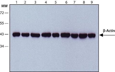
WB analysis of (1) HeLa, (2) Jurkat, (3) COS7, (4) NIH3T3, (5) PC-12, (6) RAT2, (7) CHO, (8) MDBK and (9) MDCK lysates using beta Actin antibody [AC-15] at a dilution of 1:2,000.Application
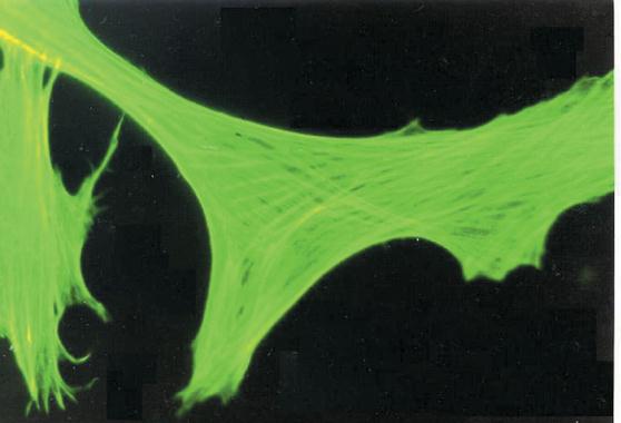
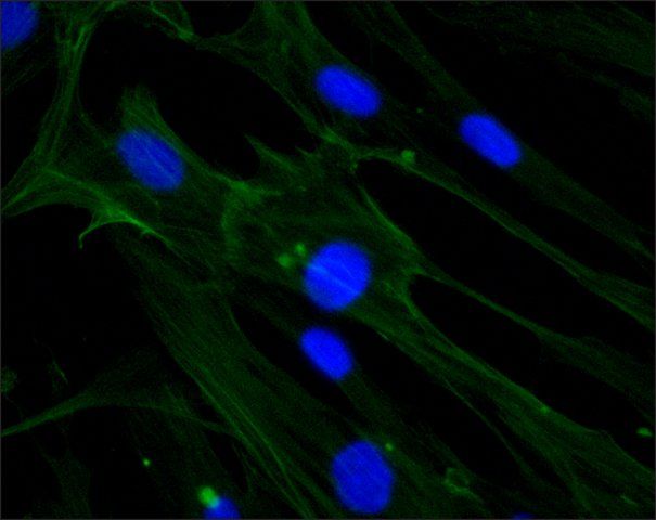
Cultured FS-11 human fibroblast cell line stained using Monoclonal Anti-β Actin, Clone No. AC-15 (Cat. No. ).Application
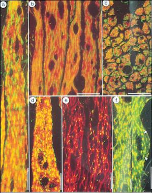
a-c. Double exposure micrographs of longitudinal, expanded (a,b) and transverse (c) semi-thin cryosections of chicken gizzard muscle double labeled with Monoclonal Anti-β-Actin, Clone No. AC-15 (Cat. No. ) (green) and polyclonal antibodies to chicken gizzard myosin (red). d-f. Longitudinal, expanded cryosections of chicken gizzard muscle double labeled with polyclonal Anti-α-Actinin (green, d, e; red, f) in combination with antibodies to chicken gizzard myosin, antibodies to chicken desmin and Monoclonal Anti-β-Actin, Clone No. AC-15 (Cat. No. ) (f, green). Bars, 10 mm.Application
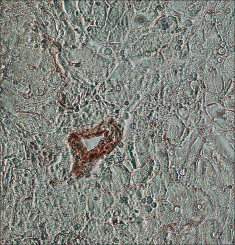
IHC-P analysis of human heart tissue using beta Actin antibody [AC-15] at a dilution of 1:200.Application
ICC/IF analysis of HS-68 cells using beta Actin antibody [AC-15] at a dilution of 1:200 (green) and DAPI (blue).Application
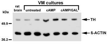
Western blot of cell lysates from whole rat brain (10 ug), untreated rat ventral mesencephalic (VM) cultures, or VM cultures treated with 1 mM db cAMP or 1 mM db cAMP + 1 uM rat galanin (2 ug) for 2 days. Lysates were separated by SDS-PAGE and immunoblottApplication
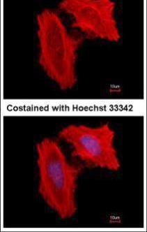
Immunofluorescence analysis of methanol-fixed HeLa, using beta Actin antibody at 1:500 dilution.Application
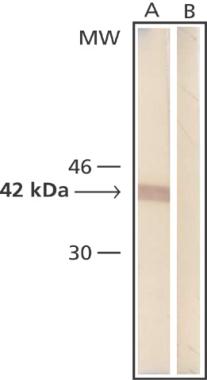
Whole cell extract of human fibroblasts was separated on SDS-PAGE and blotted with Monoclonal Anti-β-Actin (Cat No. ). The antibody was developed with Goat Anti-Mouse IgG, Peroxidase conjugate and AEC substrate. Lanes A: Antibody dilution 1:2,000 B: Negative control (only secondary antibody)| Product Name | beta Actin antibody [AC-15] |
|---|---|
| Antibody Type | Primary Antibodies |
| Product description | Mouse monoclonal [AC-15] to beta ActinBeta actin is one of six different actin isoforms which have been identified. Actins are highly conserved proteins that are involved in cell motility, structure and integrity. |
| Immunogen | Synthetic peptide: DDDIAALVIDNGSGL conjugated to KLH, corresponding to amino acids 1-14 of Xenopus laevis Actin (beta). |
| Clonality | Monoclonal |
|---|---|
| Isotype | IgG1 |
| Clone Number | AC-15 |
| Host Species | Mouse |
| Tested Applications | DotELISAIBICC/IFIHC-FIHC-PIMWB |
| WB:1:500~1:2000 IHC-P:1:50~1:200 ICC/IF:1:100~1:500 |
|
| Species Reactivity | BovineCarpCatDogDrosophilaFishGuinea PigHumanMousePigRabbitRatSheepXenopus |
| Concentration | 1mg/ml |
| Purification | Affinity purified |
| Gene Synonyms | ACTB, beta Actin, P60709, 102630, 60 |
|---|---|
| Target | beta Actin |
| Molecular Weight(MW) | 42 kDa |
| Tissue Specificity | This is an ideal reagent for use as a cell extract loading control on Western blot, Molecular weight of beta actin is 42kD. |
| Cellular Localization | Cytoplasmic |
Application
WB analysis of (1) HeLa, (2) Jurkat, (3) COS7, (4) NIH3T3, (5) PC-12, (6) RAT2, (7) CHO, (8) MDBK and (9) MDCK lysates using beta Actin antibody [AC-15] at a dilution of 1:2,000.
Application
Cultured FS-11 human fibroblast cell line stained using Monoclonal Anti-β Actin, Clone No. AC-15 (Cat. No. ).
Application
a-c. Double exposure micrographs of longitudinal, expanded (a,b) and transverse (c) semi-thin cryosections of chicken gizzard muscle double labeled with Monoclonal Anti-β-Actin, Clone No. AC-15 (Cat. No. ) (green) and polyclonal antibodies to chicken gizzard myosin (red). d-f. Longitudinal, expanded cryosections of chicken gizzard muscle double labeled with polyclonal Anti-α-Actinin (green, d, e; red, f) in combination with antibodies to chicken gizzard myosin, antibodies to chicken desmin and Monoclonal Anti-β-Actin, Clone No. AC-15 (Cat. No. ) (f, green). Bars, 10 mm.
Application
IHC-P analysis of human heart tissue using beta Actin antibody [AC-15] at a dilution of 1:200.
Application
ICC/IF analysis of HS-68 cells using beta Actin antibody [AC-15] at a dilution of 1:200 (green) and DAPI (blue).
Application
Western blot of cell lysates from whole rat brain (10 ug), untreated rat ventral mesencephalic (VM) cultures, or VM cultures treated with 1 mM db cAMP or 1 mM db cAMP + 1 uM rat galanin (2 ug) for 2 days. Lysates were separated by SDS-PAGE and immunoblott
Application
Immunofluorescence analysis of methanol-fixed HeLa, using beta Actin antibody at 1:500 dilution.
Application
Whole cell extract of human fibroblasts was separated on SDS-PAGE and blotted with Monoclonal Anti-β-Actin (Cat No. ). The antibody was developed with Goat Anti-Mouse IgG, Peroxidase conjugate and AEC substrate. Lanes A: Antibody dilution 1:2,000 B: Negative control (only secondary antibody)| Positive Control | Cultured human or chicken fibroblasts as described in Liao et alMolecular weight of beta actin is 42Kd |
|---|---|
| Application Notes | WB:1:500~1:2000 IHC-P:1:50~1:200 ICC/IF:1:100~1:500 |
| Form | Liquid |
|---|---|
| Storage Instructions | Keep as concentrated solution. Store at 4°C short term. For extended storage aliquot and store at -20°C or below. Avoid freeze-thaw cycles. |
| Storage Buffer | Ascitic fluid containing 15mM sodium azide |


