Application
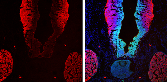
beta Tubulin 3/ TUJ1 antibody [GT11710] detects beta Tubulin 3/ TUJ1 protein by immunohistochemical analysis. Sample: Frozen sectioned E13.5 rat brain. Green: SOX2 protein stained by SOX2 antibody [N1C3] diluted at 1:250. Red: beta Tubulin 3/ TUJ1 protein stained by beta Tubulin 3/ TUJ1 antibody [GT11710] diluted at 1:250. Blue: Fluoroshield with DAPI .Application
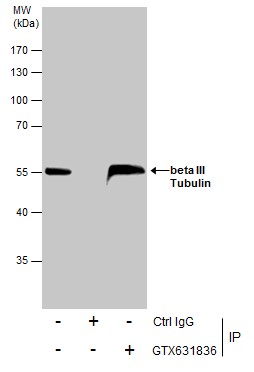
Immunoprecipitation of beta III Tubulin protein from SK-N-SH whole cell extracts using 5 μg of beta III Tubulin antibody [GT11710] . Western blot analysis was performed using beta III Tubulin antibody [GT11710] . EasyBlot anti-Mouse IgG was used as a secondary reagent.Application
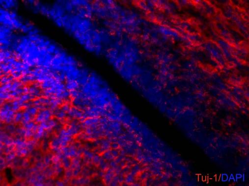
beta III Tubulin antibody [GT11710] detects beta III Tubulin proteins on embryonic mouse brain by immunohistochemical analysis. Sample:Frozen section of embryonic mouse brain (mE18.5). Red: beta III Tubulin antibody [GT11710] diluted at 1:500. Blue: DAPIApplication
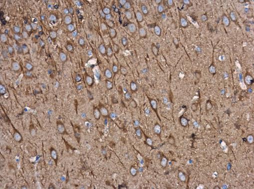
beta Tubulin 3/ TUJ1 antibody [GT11710] detects beta Tubulin 3/ TUJ1 protein at cytoplasm in rat brain by immunohistochemical analysis. Sample: Paraffin-embedded rat brain. beta Tubulin 3/ TUJ1 antibody [GT11710] diluted at 1:500.Application
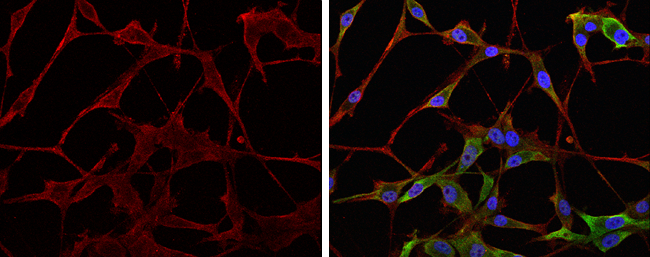
beta Tubulin 3/ TUJ1 antibody [GT11710] detects beta Tubulin 3/ TUJ1 protein at cytoskeleton by immunofluorescent analysis. Sample: U-87 MG cells were fixed in 4% paraformaldehyde at RT for 15 min. Red: beta Tubulin 3/ TUJ1 protein stained by beta Tubulin 3/ TUJ1 antibody diluted at 1:200. Green: NAGLU protein stained by NAGLU antibody [N3C1], Internal diluted at 1:200. Blue: Hoechst 33342 staining.Application
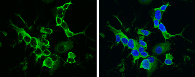
beta Tubulin 3/ TUJ1 antibody [GT11710] detects beta Tubulin 3/ TUJ1 protein at cytoskeleton by immunofluorescent analysis. Sample: SH-SY5Y cells were fixed in 4% paraformaldehyde at RT for 15 min. Green: beta Tubulin 3/ TUJ1 protein stained by beta Tubulin 3/ TUJ1 antibody [GT11710] diluted at 1:500. Blue: Hoechst 33342 staining.Application
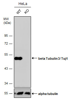
Wild-type (WT) and beta Tubulin 3/ Tuj1 knockout (KO) HeLa cell extracts (30 µg) were separated by 10% SDS-PAGE, and the membrane was blotted with beta Tubulin 3/ Tuj1 antibody [GT11710] diluted at 1:2500. The HRP-conjugated anti-mouse IgG antibody was used to detect the primary antibody, and the signal was developed with Trident ECL plus-Enhanced.Application
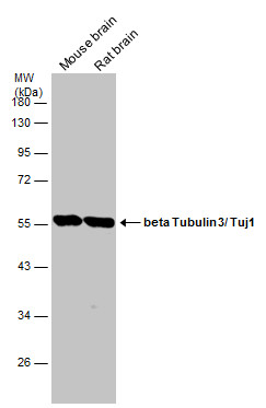
Various tissue extracts (10 µg) were separated by 10% SDS-PAGE, and the membrane was blotted with beta Tubulin 3/ Tuj1 antibody [GT11710] diluted at 1:20000. The HRP-conjugated anti-mouse IgG antibody was used to detect the primary antibody.Application
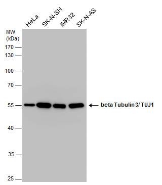
beta Tubulin 3/ TUJ1 antibody detects beta Tubulin 3/ TUJ1 protein by western blot analysis. Various whole cell extracts (20 µg) were separated by 10% SDS-PAGE, and the membrane was blotted with beta Tubulin 3/ TUJ1 antibody diluted at a dilution of 1:1000. The HRP-conjugated anti-mouse IgG antibody was used to detect the primary antibody.Application
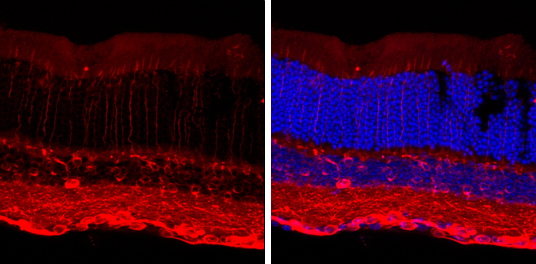
beta Tubulin 3/ TUJ1 antibody [GT11710] detects beta Tubulin 3/ TUJ1 protein by immunohistochemical analysis. Sample: Frozen sectioned adult mouse retina. Red: beta Tubulin 3/ TUJ1 protein stained by beta Tubulin 3/ TUJ1 antibody [GT11710] diluted at 1:250. Blue: Fluoroshield with DAPI .Application
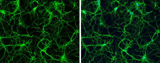
beta Tubulin 3/ TUJ1 antibody [GT11710] detects beta Tubulin 3/ TUJ1 protein expression by immunofluorescent analysis. Sample: Cultured rat E18 primary hippocampal neuron. Cells were fixed in 4% paraformaldehyde at RT for 15 min. Green: beta Tubulin 3/ TUJ1 protein stained by beta Tubulin 3/ TUJ1 antibody [GT11710] diluted at 1:250. Blue: Fluoroshield with DAPI .Application
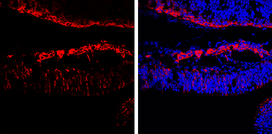
beta Tubulin 3/ TUJ1 antibody [GT11710] detects beta Tubulin 3/ TUJ1 protein by immunohistochemical analysis. Sample: Frozen sectioned E13.5 rat brain. Red: beta Tubulin 3/ TUJ1 protein stained by beta Tubulin 3/ TUJ1 antibody [GT11710] diluted at 1:250. Blue: Fluoroshield with DAPI .| Product Name | beta Tubulin 3/ Tuj1 antibody [GT11710] |
|---|---|
| Antibody Type | Primary Antibodies |
| Product description | Mouse Monoclonal antibody to beta Tubulin 3/ TUJ1 (tubulin, beta 3 class III)This gene encodes a class III member of the beta tubulin protein family. Beta tubulins are one of two core protein families (alpha and beta tubulins) that heterodimerize and assemble to form microtubules. This protein is primarily expressed in neurons and may be involved in neurogenesis and axon guidance and maintenance. Mutations in this gene are the cause of congenital fibrosis of the extraocular muscles type 3. Alternate splicing results in multiple transcript variants. A pseudogene of this gene is found on chromosome 6. |
| Immunogen | Carrier-protein conjugated synthetic peptide encompassing a sequence within the C-terminus region of human beta III Tubulin. The exact sequence is proprietary. |
| Clonality | Monoclonal |
|---|---|
| Isotype | IgG2 |
| Clone Number | M11710 |
| Host Species | Mouse |
| Tested Applications | ICC/IFIHC-FIHC-PIPWB |
| *Optimal dilutions/concentrations should be determined by the researcher.ICC/IF:1:100~1:1000 IHC:1:100~1:1000 IP:1:100~1:500 WB:1:200~1:2000 |
|
| Species Reactivity | HumanMouseRat |
| Predicted Reactivity | Chimpanzee, BovineChimpanzee (100%), Bovine (100%) |
| Concentration | 1mg/ml (Please refer to the vial label for the specific concentration) |
| Purification | Affinity purified |
| Gene Synonyms | CDCBM , CFEOM3A , TUBB4 , tubulin, beta 3 class III , beta-4 , TUBB3, TUJ1 |
|---|---|
| Target | beta Tubulin 3/ TUJ1 |
| Molecular Weight(MW) | 50 kDa (note) (2) |
| Tissue Specificity | beta III Tubulin antibody [GT11710] specifically recognizes endogenous levels of beta III Tubulin based on the immunogen sequence. |
Application
beta Tubulin 3/ TUJ1 antibody [GT11710] detects beta Tubulin 3/ TUJ1 protein by immunohistochemical analysis. Sample: Frozen sectioned E13.5 rat brain. Green: SOX2 protein stained by SOX2 antibody [N1C3] diluted at 1:250. Red: beta Tubulin 3/ TUJ1 protein stained by beta Tubulin 3/ TUJ1 antibody [GT11710] diluted at 1:250. Blue: Fluoroshield with DAPI .
Application
Immunoprecipitation of beta III Tubulin protein from SK-N-SH whole cell extracts using 5 μg of beta III Tubulin antibody [GT11710] . Western blot analysis was performed using beta III Tubulin antibody [GT11710] . EasyBlot anti-Mouse IgG was used as a secondary reagent.
Application
beta III Tubulin antibody [GT11710] detects beta III Tubulin proteins on embryonic mouse brain by immunohistochemical analysis. Sample:Frozen section of embryonic mouse brain (mE18.5). Red: beta III Tubulin antibody [GT11710] diluted at 1:500. Blue: DAPI
Application
beta Tubulin 3/ TUJ1 antibody [GT11710] detects beta Tubulin 3/ TUJ1 protein at cytoplasm in rat brain by immunohistochemical analysis. Sample: Paraffin-embedded rat brain. beta Tubulin 3/ TUJ1 antibody [GT11710] diluted at 1:500.
Application
beta Tubulin 3/ TUJ1 antibody [GT11710] detects beta Tubulin 3/ TUJ1 protein at cytoskeleton by immunofluorescent analysis. Sample: U-87 MG cells were fixed in 4% paraformaldehyde at RT for 15 min. Red: beta Tubulin 3/ TUJ1 protein stained by beta Tubulin 3/ TUJ1 antibody diluted at 1:200. Green: NAGLU protein stained by NAGLU antibody [N3C1], Internal diluted at 1:200. Blue: Hoechst 33342 staining.
Application
beta Tubulin 3/ TUJ1 antibody [GT11710] detects beta Tubulin 3/ TUJ1 protein at cytoskeleton by immunofluorescent analysis. Sample: SH-SY5Y cells were fixed in 4% paraformaldehyde at RT for 15 min. Green: beta Tubulin 3/ TUJ1 protein stained by beta Tubulin 3/ TUJ1 antibody [GT11710] diluted at 1:500. Blue: Hoechst 33342 staining.
Application
Wild-type (WT) and beta Tubulin 3/ Tuj1 knockout (KO) HeLa cell extracts (30 µg) were separated by 10% SDS-PAGE, and the membrane was blotted with beta Tubulin 3/ Tuj1 antibody [GT11710] diluted at 1:2500. The HRP-conjugated anti-mouse IgG antibody was used to detect the primary antibody, and the signal was developed with Trident ECL plus-Enhanced.
Application
Various tissue extracts (10 µg) were separated by 10% SDS-PAGE, and the membrane was blotted with beta Tubulin 3/ Tuj1 antibody [GT11710] diluted at 1:20000. The HRP-conjugated anti-mouse IgG antibody was used to detect the primary antibody.
Application
beta Tubulin 3/ TUJ1 antibody detects beta Tubulin 3/ TUJ1 protein by western blot analysis. Various whole cell extracts (20 µg) were separated by 10% SDS-PAGE, and the membrane was blotted with beta Tubulin 3/ TUJ1 antibody diluted at a dilution of 1:1000. The HRP-conjugated anti-mouse IgG antibody was used to detect the primary antibody.
Application
beta Tubulin 3/ TUJ1 antibody [GT11710] detects beta Tubulin 3/ TUJ1 protein by immunohistochemical analysis. Sample: Frozen sectioned adult mouse retina. Red: beta Tubulin 3/ TUJ1 protein stained by beta Tubulin 3/ TUJ1 antibody [GT11710] diluted at 1:250. Blue: Fluoroshield with DAPI .
Application
beta Tubulin 3/ TUJ1 antibody [GT11710] detects beta Tubulin 3/ TUJ1 protein expression by immunofluorescent analysis. Sample: Cultured rat E18 primary hippocampal neuron. Cells were fixed in 4% paraformaldehyde at RT for 15 min. Green: beta Tubulin 3/ TUJ1 protein stained by beta Tubulin 3/ TUJ1 antibody [GT11710] diluted at 1:250. Blue: Fluoroshield with DAPI .
Application
beta Tubulin 3/ TUJ1 antibody [GT11710] detects beta Tubulin 3/ TUJ1 protein by immunohistochemical analysis. Sample: Frozen sectioned E13.5 rat brain. Red: beta Tubulin 3/ TUJ1 protein stained by beta Tubulin 3/ TUJ1 antibody [GT11710] diluted at 1:250. Blue: Fluoroshield with DAPI .| Positive Control | HeLa , SK-N-SH , IMR32 , SK-N-AS , mouse brain , rat brain |
|---|---|
| Application Notes | *Optimal dilutions/concentrations should be determined by the researcher.ICC/IF:1:100~1:1000 IHC:1:100~1:1000 IP:1:100~1:500 WB:1:200~1:2000 |
| Form | Liquid |
|---|---|
| Storage Instructions | Keep as concentrated solution. Aliquot and store at -20ºC or below. Avoid multiple freeze-thaw cycles. |
| Storage Buffer | PBS |


