Application
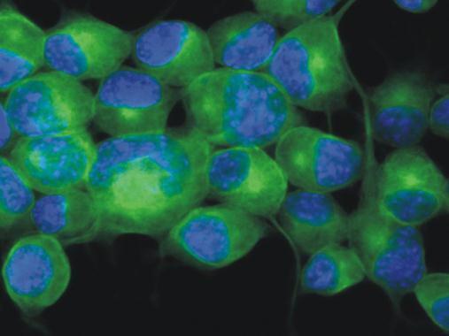
Immunofluorescence staining of Neuro2a mouse neuroblastoma cell line using anti-betaIII-tubulin ( green; 3 μg/ml). Nuclei were stained with DAPI (blue)Application
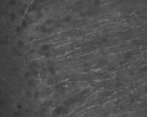
at a dilution of 1/1000, staining neuron specific beta III tubulin TU-20 (Alexa 488 secondary at 1/2000) on rat brain tissue (30m thick coronal sections) in free floating IHC (see protocol link for detailed description). Image shows labeling pf nApplication
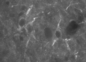
at a dilution of 1/1000, staining neuron specific beta III tubulin TU-20 (Alexa 488 secondary at 1/2000) on rat brain tissue (30m thick coronal sections) in free floating IHC (see protocol link for detailed description). Images shows cytoskeletalApplication
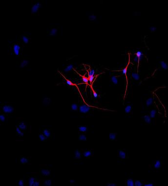
Immunofluorescence staining of P-19 mouse embryonal carcinoma cell line stimulated to neuronal differentiation by retinoic acid. 2A - Microtubules decorated with neuron-specific anti-betaIII-tubulin ( red).Application
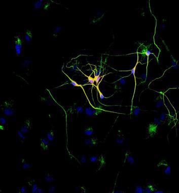
Immunofluorescence staining of P-19 mouse embryonal carcinoma cell line stimulated to neuronal differentiation by retinoic acid. 2B - Merged image of co-staining with anti-beta-tubulin . Superposition of red and green colours provided yellow staining. Nuclei were stained with DNA-binding dye (blue).| Product Name | beta Tubulin 3/ Tuj1 antibody [TU-20] |
|---|---|
| Antibody Type | Primary Antibodies |
| Product description | Mouse monoclonal antibody [TU-20] to beta III TubulinThis gene encodes a class III member of the beta tubulin protein family. Beta tubulins are one of two core protein families (alpha and beta tubulins) that heterodimerize and assemble to form microtubules. This protein is primarily expressed in neurons and may be involved in neurogenesis and axon guidance and maintenance. Mutations in this gene are the cause of congenital fibrosis of the extraocular muscles type 3. Alternate splicing results in multiple transcript variants. A pseudogene of this gene is found on chromosome 6. [provided by RefSeq] |
| Immunogen | Synthetic peptide: ESESQGPK conjugated to malemide activated KLH, corresponding to amino acids 441-448 of Human Tubulin. |
| Clonality | Monoclonal |
|---|---|
| Isotype | IgG1 |
| Clone Number | TU-20 |
| Host Species | Mouse |
| Tested Applications | FACSICC/IFIHCWB |
| Western Blotting | |
| Species Reactivity | HumanMousePigRatBaboonCarpHamsterMammalian speciesBovine |
| Concentration | 1mg/ml |
| Purification | Affinity purified |
| Gene Synonyms | CFEOM3A, TUBB4, beta-4, beta4 |
|---|---|
| Target | beta III Tubulin |
| Tissue Specificity | Class III beta-tubulin specific for neurones. |
Application
Immunofluorescence staining of Neuro2a mouse neuroblastoma cell line using anti-betaIII-tubulin ( green; 3 μg/ml). Nuclei were stained with DAPI (blue)
Application
at a dilution of 1/1000, staining neuron specific beta III tubulin TU-20 (Alexa 488 secondary at 1/2000) on rat brain tissue (30m thick coronal sections) in free floating IHC (see protocol link for detailed description). Image shows labeling pf n
Application
at a dilution of 1/1000, staining neuron specific beta III tubulin TU-20 (Alexa 488 secondary at 1/2000) on rat brain tissue (30m thick coronal sections) in free floating IHC (see protocol link for detailed description). Images shows cytoskeletal
Application
Immunofluorescence staining of P-19 mouse embryonal carcinoma cell line stimulated to neuronal differentiation by retinoic acid. 2A - Microtubules decorated with neuron-specific anti-betaIII-tubulin ( red).
Application
Immunofluorescence staining of P-19 mouse embryonal carcinoma cell line stimulated to neuronal differentiation by retinoic acid. 2B - Merged image of co-staining with anti-beta-tubulin . Superposition of red and green colours provided yellow staining. Nuclei were stained with DNA-binding dye (blue).| Application Notes | Western Blotting |
|---|
| Form | Liquid |
|---|---|
| Storage Instructions | Keep as concentrated solution. Store at 4°C short term. For extended storage aliquot and store at -20°C or below. Avoid freeze-thaw cycles. |
| Storage Buffer | Phosphate buffered saline pH 7.4 containing 15mM sodium azide |


