Application
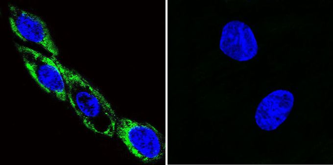
Immunofluorescent analysis of Thr286-phosphorylated CaM Kinase II in C6 cells. Thr286-phosphorylated CaM Kinase II staining (green) and nuclei with DAPI (blue) is shown. Cells were grown on chamber slides and fixed with formaldehyde prior to staining. Cells were probed without (control) or with CaM Kinase II (phospho Thr286) antibody [22B1] at a dilution of 1:100 over night at 4 ?C, washed with PBS and incubated with a proper secondary antibody. Images were taken at 60X magnification.| Product Name | CaMKII alpha (phospho Thr286) antibody |
|---|---|
| Antibody Type | Primary Antibodies |
| Product description | Mouse monoclonal antibody [22B1] to Thr286-phosphorylated CaM Kinase II |
| Immunogen | Synthetic peptide corresponding to residues M(281) H R Q E T(p) V D C L K K F N(294) of rat CaM Kinase II protein. |
| Modification | p-Thr286 |
| Clonality | Monoclonal |
|---|---|
| Isotype | IgG1 |
| Clone Number | 22B1 |
| Host Species | Mouse |
| Tested Applications | ELISAFACSICC/IFIHCIPWB |
| ICC/IF: 1:50 - 1:1000: |
|
| Species Reactivity | HumanMouseRatRabbit |
| Concentration | 1mg/ml (Please refer to the vial label for the specific concentration) |
| Purification | Protein G |
| Gene Synonyms | PKCCD calcium/calmodulin-dependent protein kinase II alpha PK2CDD Camk2a |
|---|---|
| Target | Phospho-CaM Kinase II pThr286 |
| Tissue Specificity | This antibody has been shown to detect the phosphorylated alpha subunit CaM kinase II only. |
Application
Immunofluorescent analysis of Thr286-phosphorylated CaM Kinase II in C6 cells. Thr286-phosphorylated CaM Kinase II staining (green) and nuclei with DAPI (blue) is shown. Cells were grown on chamber slides and fixed with formaldehyde prior to staining. Cells were probed without (control) or with CaM Kinase II (phospho Thr286) antibody [22B1] at a dilution of 1:100 over night at 4 ?C, washed with PBS and incubated with a proper secondary antibody. Images were taken at 60X magnification.| Application Notes | ICC/IF: 1:50 - 1:1000: |
|---|
| Form | Liquid |
|---|---|
| Storage Instructions | -20° C, Avoid Freeze/Thaw Cycles |
| Storage Buffer | PBS containing 0.05 % sodium azide |


