Application
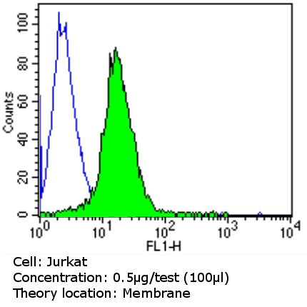
Flow cytometry analysis of CD18 in Jurkat cells (green) compared to an isotype control (blue). Cells were harvested, adjusted to a concentration of 1-5x10^6 cells/ml, fixed with 2% paraformaldehyde and washed with PBS. Cells were blocked with a 2% solution of BSA-PBS for 30 min at room temperature and incubated with CD18/ITGB2 antibody [TS1/18] at a dilution of 0.5 ug/test for 60 min at room temperature. Cells were then incubated in a proper secondary antibody and re-suspended in PBS for FACS analysis.Application
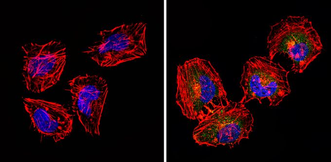
Immunofluorescent analysis of CD18 (green) in HeLa cells (right) compared to a negative control without primary antibody (left). Formalin-fixed cells were permeabilized with 0.1% Triton X-100 in TBS for 5-10 minutes and blocked with 3% BSA-PBS for 30 minutes at room temperature. Cells were probed with CD18/ITGB2 antibody [TS1/18] in 3% BSA-PBS at a dilution of 1:20 and incubated overnight at 4°C in a humidified chamber. Cells were washed with PBST and incubated with a proper secondary antibody in PBS at room temperature in the dark. F-actin (red) was stained with a flourescent red phalloidin and nuclei (blue) were stained with Hoechst or DAPI. Images were taken at a magnification of 60x.| Product Name | CD18 antibody |
|---|---|
| Antibody Type | Primary Antibodies |
| Product description | Mouse Monoclonal antibody [TS1/18] to CD18The product of this gene belongs to the integrin beta chain family of proteins. Integrins are integral cell-surface proteins composed of an alpha chain and a beta chain. This gene encodes the integrin beta chain beta 2. A given chain may combine with multiple partners resulting in different integrins. For example, beta 2 combines with the alpha L chain to form the integrin LFA-1, and combines with the alpha M chain to form the integrin Mac-1. Integrins are known to participate in cell adhesion as well as cell-surface mediated signalling. Defects in this gene are the cause of leukocyte adhesion deficiency type I (LAD1). Two transcript variants encoding the same protein have been identified for this gene. [provided by RefSeq, Jul 2008] |
| Immunogen | Human Beta 2 Integrin |
| Clonality | Monoclonal |
|---|---|
| Isotype | IgG1 |
| Clone Number | TS1/18 |
| Host Species | Mouse |
| Tested Applications | BlockingFACSICC/IFIHCInhibitionWB |
| ICC/IF: 1:10 - 1:100: |
|
| Species Reactivity | HumanMouseHamster |
| Concentration | 1mg/ml |
| Purification | Protein G |
| Gene Synonyms | MF17 integrin beta 2 (complement component 3 receptor 3 and 4 subunit) ITGB2 LCAMB MFI7 LAD CD18 MAC-1 LFA-1 |
|---|---|
| Target | CD18 |
Application
Flow cytometry analysis of CD18 in Jurkat cells (green) compared to an isotype control (blue). Cells were harvested, adjusted to a concentration of 1-5x10^6 cells/ml, fixed with 2% paraformaldehyde and washed with PBS. Cells were blocked with a 2% solution of BSA-PBS for 30 min at room temperature and incubated with CD18/ITGB2 antibody [TS1/18] at a dilution of 0.5 ug/test for 60 min at room temperature. Cells were then incubated in a proper secondary antibody and re-suspended in PBS for FACS analysis.
Application
Immunofluorescent analysis of CD18 (green) in HeLa cells (right) compared to a negative control without primary antibody (left). Formalin-fixed cells were permeabilized with 0.1% Triton X-100 in TBS for 5-10 minutes and blocked with 3% BSA-PBS for 30 minutes at room temperature. Cells were probed with CD18/ITGB2 antibody [TS1/18] in 3% BSA-PBS at a dilution of 1:20 and incubated overnight at 4°C in a humidified chamber. Cells were washed with PBST and incubated with a proper secondary antibody in PBS at room temperature in the dark. F-actin (red) was stained with a flourescent red phalloidin and nuclei (blue) were stained with Hoechst or DAPI. Images were taken at a magnification of 60x.| Application Notes | ICC/IF: 1:10 - 1:100: |
|---|
| Form | Liquid |
|---|---|
| Storage Instructions | -20°C |
| Storage Buffer | PBS |


