Application
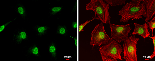
CDC25C antibody detects CDC25C protein at nucleus by immunofluorescent analysis. Sample: HeLa cells were fixed in 4% paraformaldehyde at RT for 15 min. Green: CDC25C protein stained by CDC25C antibody diluted at 1:500. Red: phalloidin, a cytoskeleton marker, diluted at 1:200. Scale bar = 10 μm.Application
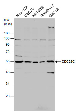
Various whole cell extracts (30 μg) were separated by 10% SDS-PAGE, and the membrane was blotted with CDC25C antibody diluted at 1:500.Application
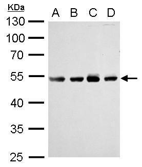
CDC25C antibody detects CDC25C protein by Western blot analysis. A. 30 µg Jurkat whole cell lysate/extract B. 30 µg Raji whole cell lysate/extract C. 30 µg K562 whole cell lysate/extract D. 30 µg NCI-H929 whole cell lysate/extract 10 % SDS-PAGE CDC25C antibody dilution: 1:1000Application
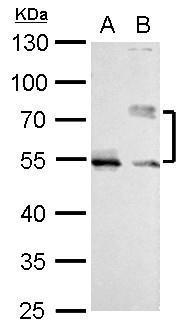
CDC25C antibody detects CDC25C protein by Western blot analysis. A. 30 µg HeLa whole cell lysate/extract (untreated) B. 30 µg HeLa whole cell lysate/extract (100ng/ml Nocodazole treatment for 24hr) 10 % SDS-PAGE CDC25C antibody dilution: 1:1000| Product Name | CDC25C antibody |
|---|---|
| Antibody Type | Primary Antibodies |
| Product description | Rabbit Polyclonal antibody to CDC25C (cell division cycle 25 homolog C (S. pombe))This gene is highly conserved during evolution and it plays a key role in the regulation of cell division. The encoded protein is a tyrosine phosphatase and belongs to the Cdc25 phosphatase family. It directs dephosphorylation of cyclin B-bound CDC2 and triggers entry into mitosis. It is also thought to suppress p53-induced growth arrest. Multiple alternatively spliced transcript variants of this gene have been described, however, the full-length nature of many of them is not known. [provided by RefSeq] |
| Immunogen | Carrier-protein conjugated synthetic peptide encompassing a sequence within the center region of human CDC25C. The exact sequence is proprietary. |
| Clonality | Polyclonal |
|---|---|
| Isotype | IgG |
| Host Species | Rabbit |
| Tested Applications | ICC/IFWB |
| *Optimal dilutions/concentrations should be determined by the researcher.
: |
|
| Species Reactivity | HumanMouseRat |
| Predicted Reactivity | ChimpanzeeChimpanzee (100%) |
| Concentration | 1mg/ml (Please refer to the vial label for the specific concentration) |
| Purification | Affinity purified |
| Gene Synonyms | CDC25 antibody CDC25C antibody mitosis inducer CDC25 antibody dual specificity phosphatase CDC25C antibody M-phase inducer phosphatase 3 antibody cell division cycle 25C antibody protein phosphatase 1 regulatory subunit 60 antibody phosphotyrosine |
|---|---|
| Molecular Weight(MW) | 53 kDa (note) (5) |
| Cellular Localization | Nucleus |
Application
CDC25C antibody detects CDC25C protein at nucleus by immunofluorescent analysis. Sample: HeLa cells were fixed in 4% paraformaldehyde at RT for 15 min. Green: CDC25C protein stained by CDC25C antibody diluted at 1:500. Red: phalloidin, a cytoskeleton marker, diluted at 1:200. Scale bar = 10 μm.
Application
Various whole cell extracts (30 μg) were separated by 10% SDS-PAGE, and the membrane was blotted with CDC25C antibody diluted at 1:500.
Application
CDC25C antibody detects CDC25C protein by Western blot analysis. A. 30 µg Jurkat whole cell lysate/extract B. 30 µg Raji whole cell lysate/extract C. 30 µg K562 whole cell lysate/extract D. 30 µg NCI-H929 whole cell lysate/extract 10 % SDS-PAGE CDC25C antibody dilution: 1:1000
Application
CDC25C antibody detects CDC25C protein by Western blot analysis. A. 30 µg HeLa whole cell lysate/extract (untreated) B. 30 µg HeLa whole cell lysate/extract (100ng/ml Nocodazole treatment for 24hr) 10 % SDS-PAGE CDC25C antibody dilution: 1:1000| Positive Control | Jurkat , Raji , K562 , NCI-H929 , HeLa (untreated) , HeLa (100ng/ml Nocodazole treatment for 24hr) , Neuro 2A , C8D30 , NIH-3T3 , Raw264.7 , C2C12 , PC-12 , Rat2 |
|---|---|
| Application Notes | *Optimal dilutions/concentrations should be determined by the researcher.
: |
| Form | Liquid |
|---|---|
| Storage Instructions | Keep as concentrated solution. Aliquot and store at -20ºC or below. Avoid multiple freeze-thaw cycles. |
| Storage Buffer | 1XPBS, 20% Glycerol (pH7). 0.01% Thimerosal was added as a preservative. |


