Application
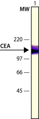
WB ImageCEA preparation extract was separated on SDS-PAGE and probed with Monoclonal AB To CEA (MO)CL:C6G9 (PREPK) . The antibody was developed with Alk Phos APA Mouse Fab ads HIgG and a NBT/BCIP substrate. Antibody dilution 1/1500| Product Name | CEA antibody |
|---|---|
| Antibody Type | Primary Antibodies |
| Product description | Mouse monoclonal [C6G9] to Carcino Embryonic Antigen CEACarcino Embryonic Antigen (CEA) is synthesized during development in the fetal gut, and is re expressed in increased amounts in intestinal carcinomas and several other tumors. Antibodies to CEA are useful in identifying the origin of various metastatic adenocarcinomas and in distinguishing pulmonary adenocarcinomas (60 to 70% are CEA+) from pleural mesotheliomas (rarely or weakly CEA+). |
| Immunogen | CEA isolated from a human colon adenocarcinoma cell line. |
| Clonality | Monoclonal |
|---|---|
| Isotype | IgG1 |
| Clone Number | C6G9 |
| Host Species | Mouse |
| Tested Applications | DotIHC-PWB |
| Dot: Use at a dilution of 1/8000. IHC-P: Use at a dilution of 1/8000. Perform enzymatic antigen retrieval before commencing with IHC staining protocol. WB: Use at a dilution of 1/8000. Predicted molecular weight: 180 kDa. There is no cross reactivity with nonspecific cross-reacting antigen (NCA) present in granulocytes by immunoblotting, flow cytometry or immunohistochemical techniques. The antibody recognizes an epitope that is resistant to 30 minute oxidation by 1% sodium periodate solution in routine formalin-fixed, paraffin-embedded tissue sections. Enzymatic predigestion with proteolytic enzymes enhances immunohistochemical staining of formalinfixed, paraffin-embedded tissue sections with the antibody. Optimal dilutions/concentrations should be determined by the end user.: |
|
| Species Reactivity | Human |
| Concentration | 1mg/ml |
| Purification | Affinity purified |
| Gene Synonyms | P06731 CEACAM5 114890 CD66e 1048 DKFZp781M2392 CEA |
|---|---|
| Target | CEA |
| Tissue Specificity | Monoclonal Anti-Human Carcinoembryonic Antigen (CEA) reacts specifically with human CEA (CD66e, 180 kDa) from several types of malignant tissues including colorectal, lung and breast tumors. It also stains medullary carcinomas of the thyroid. The product reacts strongly with the cell surface and cytoplasm of malignant glands in colorectal adenocarcinomas. A weak reactivity is seen with normal colon mucosa and occasionally with bile canalicular and pancreatic acinar cells, but not with other tested normal tissues including term placenta. |
| Cellular Localization | Plasma membrane |
Application
WB ImageCEA preparation extract was separated on SDS-PAGE and probed with Monoclonal AB To CEA (MO)CL:C6G9 (PREPK) . The antibody was developed with Alk Phos APA Mouse Fab ads HIgG and a NBT/BCIP substrate. Antibody dilution 1/1500| Application Notes | Dot: Use at a dilution of 1/8000. IHC-P: Use at a dilution of 1/8000. Perform enzymatic antigen retrieval before commencing with IHC staining protocol. WB: Use at a dilution of 1/8000. Predicted molecular weight: 180 kDa. There is no cross reactivity with nonspecific cross-reacting antigen (NCA) present in granulocytes by immunoblotting, flow cytometry or immunohistochemical techniques. The antibody recognizes an epitope that is resistant to 30 minute oxidation by 1% sodium periodate solution in routine formalin-fixed, paraffin-embedded tissue sections. Enzymatic predigestion with proteolytic enzymes enhances immunohistochemical staining of formalinfixed, paraffin-embedded tissue sections with the antibody. Optimal dilutions/concentrations should be determined by the end user.: |
|---|
| Form | Liquid |
|---|---|
| Storage Instructions | Keep as concentrated solution. Store at 4ºC short term. For extended storage aliquot and store at -20ºC or below. Avoid freeze-thaw cycles. |
| Storage Buffer | Preservative: 0.1% Sodium Azide |


